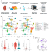
Renal Single-Cell RNA Sequencing and Digital Cytometry in Dogs with X-Linked Hereditary Nephropathy
- PMID: 40723524
- PMCID: PMC12291704
- DOI: 10.3390/ani15142061
Renal Single-Cell RNA Sequencing and Digital Cytometry in Dogs with X-Linked Hereditary Nephropathy
Abstract
Chronic kidney disease (CKD) significantly affects canine health, but the precise cellular mechanisms of this condition remain elusive. In this study, we used single-cell RNA sequencing (scRNA-seq) to profile renal cellular gene expression in a canine model of X-linked hereditary nephropathy (XLHN). Dogs with this condition exhibit juvenile-onset CKD similar to that seen in human Alport syndrome. Post-mortem renal cortical tissues from an affected male dog and a heterozygous female dog were processed to obtain single-cell suspensions. In total, we recovered up to 13,190 cells and identified 11 cell types, including major kidney cells and immune cells. Differential gene expression analysis comparing the affected male and heterozygous female dogs identified cell-type specific pathways that differed in a subpopulation of proximal tubule cells. These pathways included the integrin signaling pathway and the pathway for inflammation mediated by chemokine and cytokine signaling. Additionally, using machine learning-empowered digital cytometry, we deconvolved bulk mRNA-seq data from a previous canine study, revealing changes in cell type proportions across CKD stages. These results underline the utility of single-cell methodologies and digital cytometry in veterinary nephrology.
Keywords: Alport syndrome; RNA-seq; X-linked hereditary nephropathy; digital cytometry; dog; kidney; scRNA-seq.
Conflict of interest statement
Author Daniel Osorio was employed by the company Qiagen Digital Insight. The remaining authors declare that the research was conducted in the absence of any commercial or financial relationships that could be construed as a potential conflict of interest.
Figures


Similar articles
-
Interleukin (IL)-34 promotes the inflammatory role of IL-1β-producing myeloid cells in pemphigus lesions.Br J Dermatol. 2025 Jul 17;193(2):287-297. doi: 10.1093/bjd/ljaf130. Br J Dermatol. 2025. PMID: 40203120
-
Comprehensive single-cell chromatin and transcriptomic profiling of peripheral immune cells in nonsegmental vitiligo.Br J Dermatol. 2025 Jun 20;193(1):115-124. doi: 10.1093/bjd/ljaf041. Br J Dermatol. 2025. PMID: 39888372
-
Systemic pharmacological treatments for chronic plaque psoriasis: a network meta-analysis.Cochrane Database Syst Rev. 2021 Apr 19;4(4):CD011535. doi: 10.1002/14651858.CD011535.pub4. Cochrane Database Syst Rev. 2021. Update in: Cochrane Database Syst Rev. 2022 May 23;5:CD011535. doi: 10.1002/14651858.CD011535.pub5. PMID: 33871055 Free PMC article. Updated.
-
Chondrodystrophic Dogs as a Preclinical Large Animal Model of Discogenic Back Pain.JOR Spine. 2025 Jul 14;8(3):e70082. doi: 10.1002/jsp2.70082. eCollection 2025 Sep. JOR Spine. 2025. PMID: 40662113 Free PMC article.
-
Angiotensin-converting enzyme inhibitors and angiotensin receptor blockers for adults with early (stage 1 to 3) non-diabetic chronic kidney disease.Cochrane Database Syst Rev. 2011 Oct 5;(10):CD007751. doi: 10.1002/14651858.CD007751.pub2. Cochrane Database Syst Rev. 2011. Update in: Cochrane Database Syst Rev. 2023 Jul 19;7:CD007751. doi: 10.1002/14651858.CD007751.pub3. PMID: 21975774 Updated.
References
-
- Zheng K., Thorner P.S., Marrano P., Baumal R., McInnes R.R. Canine X Chromosome-Linked Hereditary Nephritis: A Genetic Model for Human X-Linked Hereditary Nephritis Resulting from a Single Base Mutation in the Gene Encoding the Alpha 5 Chain of Collagen Type IV. Proc. Natl. Acad. Sci. USA. 1994;91:3989–3993. doi: 10.1073/pnas.91.9.3989. - DOI - PMC - PubMed
Grants and funding
LinkOut - more resources
Full Text Sources

