Review
. 2025 Jun 24;15(7):925.
doi: 10.3390/biom15070925.

Female Reproductive Tract Organoids: Applications from Physiology to Pathology

Affiliations
Review

Female Reproductive Tract Organoids: Applications from Physiology to Pathology

Xinyu Wang et al. Biomolecules. .

Abstract

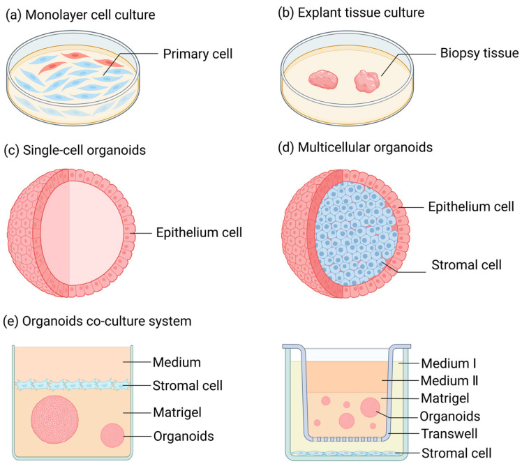
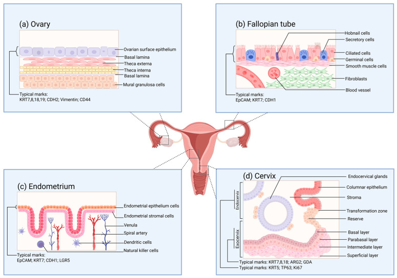
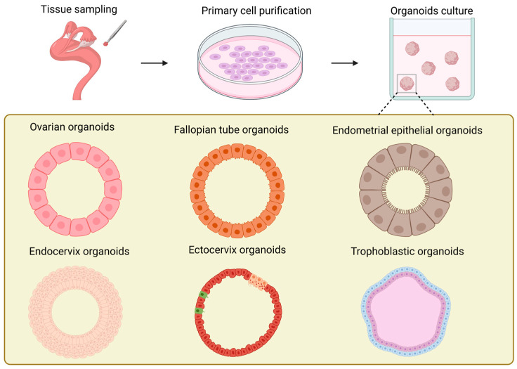
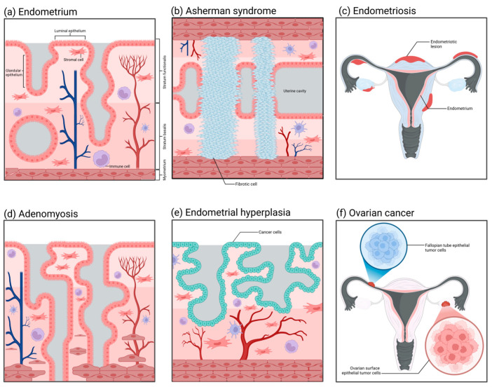
The female reproductive tract (FRT) serves as the core of human reproduction, and its health is directly related to population quantity and family happiness. The high incidence rate of female reproductive tract diseases globally poses a severe threat to women's health. Nevertheless, the exploration of its physiological functions and pathological mechanisms still lacks satisfactory research models. Organoids, as an emerging technology, not only circumvent numerous ethical issues existing in in vivo experiments but also precisely replicate the morphological structure and characteristics of the simulated tissues. The purpose of this article is to summarize the basic paradigms of organoid establishment and their applications in female reproductive research. Specifically, this article summarizes the cell sources, extracellular scaffolds, and culture media used in the establishment of organoids. It also describes the applications and future development prospects of female reproductive tract organoids established in current research in physiological and pathological studies. The importance of organoid technology in the female reproductive tract research cannot be ignored. It has opened up new avenues for research in this field and greatly promoted the exploration of female reproductive health and disease mechanisms.

Keywords: female reproductive tract; hydrogel; organoid; reproductive disorder; reproductive physiology.


Conflict of interest statement

The authors declare no conflicts of interest.

Figures

Figure 1
Figure 1
Research methods for female reproductive tract diseases. (a) Monolayer cell culture: A method of cell culture in animal cells where cells rapidly spread and start mitosis once adhered to the surface, gradually forming a dense monolayer of cells. (b) Tissue block culture: A method of culturing where freshly isolated, highly viable tissue is cut into small pieces and inoculated into culture flasks for growth. (c) Single-cell organoids: They are derived from a single type of stem cell or purified primary cells. Prior to organoid culture, these initial cells exist in either an undifferentiated state (stem cells) or a highly differentiated state (mature somatic cells, such as endometrial epithelial cells), featuring a singular and stable transcriptional level. (d) Multicellular organoids: Cell aggregates consisting of multiple cell types. These refer to multiple types of cells or a variety of highly differentiated primary somatic cells that are obtained by inducing the differentiation of stem cells through in vitro culture techniques prior to the initiation of organoid culture. (e) Organoid co-culture system: A system where different types of organoids and cells are co-cultured to simulate the interactions between different tissues or organs in vivo.
Figure 2
Figure 2
The surface structure of the functional layers of various parts of the female reproductive tract under normal conditions. (a) Schematic diagram of the tissue structure of the functional layer on the surface of the ovary. The cell markers of the ovarian surface epithelium are also provided in the figure. These markers are of great reference significance for verifying whether the organoids are constructed successfully. (b) Schematic diagram of the tissue structure of the functional layer on the inner side of the fallopian tube. The cell markers of the inner mucosal epithelium of the fallopian tube are also provided in the figure. (c) Schematic diagram of the tissue structure of the functional layer on the surface of the endometrium. The cell markers of the endometrial epithelium are also provided in the figure. (d) Schematic diagram of the tissue structure of the functional layer on the surface of the cervix. The cell markers of the endocervix and the exocervix are also provided in the figure.
Figure 3
Figure 3
Schematic illustration of the generation and models of organoids in various female reproductive tracts.
Figure 4
Figure 4
The histopathological structures of common diseases in the female reproductive tract. (a) Normal endometrium: It is divided into the basal layer and the functional layer, containing epithelial cells, stromal cells, blood vessels, and immune cells. (b) Intrauterine adhesions (Asherman syndrome): Adhesions form within the uterine cavity, with the functional layer epithelium replaced by fibrous tissue, lacking blood vessels and glands. (c) Endometriosis: Endometrial-like tissue grows outside of the uterus. (d) Adenomyosis: Endometrial glands and stroma invade the myometrium, causing the uterus to enlarge. (e) Endometrial hyperplasia: Abnormal proliferation of endometrial epithelial cells, which may develop into endometrial cancer. (f) Ovarian cancer: Abnormal proliferation of ovarian cells forms a tumor, which can spread to other areas.
Figure 5
Figure 5
The future applications and prospects of reproductive tract organoids. (a) Organoid models can be used to study the physiology and pathology of the reproductive tract and also for genetic engineering, such as CRISPR/Cas9 gene editing or lentiviral transfection. (b) The basic procedure for establishing organoids. (c) Organoids can be used to test drug responses, accelerate drug development and screening, and assist patients with rare diseases in gene therapy.

Similar articles

References

    1. Suarez S.S., Pacey A.A. Sperm transport in the female reproductive tract. Hum. Reprod. Update. 2006;12:23–37. doi: 10.1093/humupd/dmi047. - DOI - PubMed
    1. Roy A., Matzuk M.M. Reproductive tract function and dysfunction in women. Nat. Rev. Endocrinol. 2011;7:517–525. doi: 10.1038/nrendo.2011.79. - DOI - PubMed
    1. Dalton L. Understanding the Female Reproductive Tract’s Role in Fertilization. ACS Cent. Sci. 2022;8:3–6. doi: 10.1021/acscentsci.1c01595. - DOI - PMC - PubMed
    1. Bloise E., Ciarmela P., Dela Cruz C., Luisi S., Petraglia F., Reis F.M. Activin A in Mammalian Physiology. Physiol. Rev. 2019;99:739–780. doi: 10.1152/physrev.00002.2018. - DOI - PubMed
    1. Christensen A., Bentley G.E., Cabrera R., Ortega H.H., Perfito N., Wu T.J., Micevych P. Hormonal regulation of female reproduction. Horm. Metab. Res. = Horm.-Und Stoffwechselforschung = Horm. Metab. 2012;44:587–591. doi: 10.1055/s-0032-1306301. - DOI - PMC - PubMed

LinkOut - more resources