. 2025 Jun 27;15(7):939.
doi: 10.3390/biom15070939.

β-Secosterol, an Oxyphytosterol Produced Through the Reaction of β-Sitosterol with Ozone, Demonstrates Different Cytotoxic Effects on BRL-3A and HTC Cells

Affiliations

β-Secosterol, an Oxyphytosterol Produced Through the Reaction of β-Sitosterol with Ozone, Demonstrates Different Cytotoxic Effects on BRL-3A and HTC Cells

Bianca S Takayasu et al. Biomolecules. .

Abstract

Sitosterol (Sito) is a phytosterol with bioactive properties, including reducing atherosclerosis risk and anti-inflammatory and antitumoral effects. However, it can be oxidized by reactive oxygen species such as ozone (O3), producing oxyphytosterols with harmful effects such as cytotoxicity, oxidative stress, and proatherogenicity. Ozone, a strong oxidant and common pollutant, can alter plant steroid compounds, raising concerns about dietary oxyphytosterol intake. Studies identify β-Secosterol (βSec) as the primary ozone-derived oxyphytosterol from Sito, exhibiting cytotoxic effects on HepG2 human liver tumor cells. This study investigated βSec's biological effects on two rat liver cell lines: BRL-3A (immortalized) and HTC (tumoral), examining cell death, cell cycle progression, morphology, and cytoskeleton organization. While Sito influenced cell metabolic activity without affecting cell survival or morphology, βSec demonstrated significant cytotoxicity in both cell lines. It induced G0/G1 cell cycle arrest and disrupted cytoskeleton organization, with different implications: BRL-3A cells showed persistent cytoskeletal changes potentially linked to tumor induction, while HTC cells displayed chemoresistance, restoring cytoskeletal integrity and enhancing metastatic potential. These findings reveal βSec's complex, context-dependent effects, suggesting it may promote tumor-like behavior in non-tumoral cells and resistance mechanisms in cancer cells, contributing to understanding oxyphytosterols' implications for physiological and pathological conditions.

Keywords: cell cycle; cytoskeleton; oxyphytosterols; ozone; sitosterol; β-secosterol.


Conflict of interest statement

The authors declare no conflicts of interest.

Figures

Figure 1
Figure 1
β-Secosterol (βSec) formation from Sitosterol (Sito) ozonization (for synthesis and characterization details, see Martins et al., 2020 [19]).
Figure 2
Figure 2
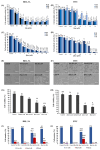
Cell metabolic activity and viability. Cell metabolic activity was measured by MTT assay, BRL-3A (A) and HTC (B) cells exposed to 0.05 µM to 20 µM Sito for 24 h, 48 h, and 72 h; BRL-3A (C) and HTC (D) cells exposed to 0.1 µM to 20 µM βSec for 24 h, 48 h, and 72 h. Data expressed as mean ± SEM of the relative percentage of the control. Statistical analysis was performed by ANOVA-Dunnett’s test for multiple comparisons versus the control (* p < 0.5, ** p < 0.01, *** p < 0.001, **** p < 0.0001). Observation of cell morphology in phase microscopy of BRL-3A (E) and HTC (F) cells and cell viability measured by trypan blue exclusion assay (G and H, respectively) after exposure of cells to βSito (0.1 and 0.2 µM for BRL-3A; 0.2 and 0.5 µM for HTC) and βSec (2 and 4 µM for BRL-3A; 1 and 2 µM for HTC) for 48 h. Counting of the number of live cells (Hoechst 33342), dead cells (propidium iodide), and mitosis rate after treatment for 48 h (Trat) and after 48 h recovery (Rec) of BRL-3A cells (I) to 4 μM βSec or HTC cells (J) to 2 μM βSec. Data are expressed as the mean ± SEM of the percentage relative to the control of three independent experiments with three replicates for each sample. Statistical analysis was performed using ANOVA-Dunnett’s test for multiple comparisons versus control (* p < 0.5, ** p < 0.01, *** p < 0.001, **** p < 0.0001). Scale bar: 200 µm (E,F).
Figure 3
Figure 3
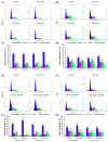
Distribution of cell cycle phases. Flow cytometry distribution of cell cycle phases of BRL-3A (A,C) and HTC cells (B,D) after treatment with Sito (0.1 and 0.2 µM for BRL-3A; 0.2 and 0.5 µM for HTC) and βSec (2 µM for BRL-3A; 1 µM for HTC) for 48 h. BRL-3A (4 µM) (E,G) and HTC (2 µM) (F,H) cells were treated with βSec for 48 h and recovered for 48 h. Data are presented as the mean ± SEM of the relative percentage compared to the control, based on three independent experiments performed in duplicate for each sample. Statistical analysis was conducted using ANOVA-Dunnett’s test for multiple comparisons vs. control (* p < 0.05, ** p < 0.01, **** p < 0.0001).
Figure 4
Figure 4
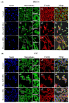
Morphology and cytoskeletal organization of BRL-3A (A) and HTC (B) cells. Fluorescence microscopy images of control (aa3) and after 48 h treatment (bb3) with Sito 0.2 µM (BRL-3A) and 0.5 µM (HTC), submitted to immunofluorescence reaction with antibodies against α and β-tubulin plus anti-mouse secondary antibodies conjugated to Alexa Fluor 488 (green). Microfilaments of f-actin were stained with phalloidin conjugated to Alexa Fluor 555 (red). Nuclei were stained with DAPI (blue). Scale bar: 30 µm (A); 25 µm (B).
Figure 5
Figure 5
Morphology and cytoskeleton organization of BRL-3A (A) and HTC (B) cells. Fluorescence microscopy images of control (aa3,a’,a”), after 48 h of treatment with 2 or 4 µM βSec (bb3,b’,b”), and after 48 h of recovery (cc3,c’,c”). Cells were subjected to immunofluorescence staining with antibodies against α- and β-tubulin, plus secondary anti-mouse antibodies conjugated to Alexa Fluor 488 (green). F-actin filaments were stained with phalloidin conjugated to Alexa Fluor 555 (red), and nuclei were stained with DAPI (blue). Scale bar: 25 µm.
Figure 6
Figure 6
Analysis of focal adhesion in BRL-3A and HTC cells. Analysis of focal adhesion in BRL-3A (A) and HTC (B) cells. Fluorescence microscopy images of control (aa3), after 48 h of treatment with βSec 4 µM for BRL-3A and 2 µM for HTC (bb3), and after 48 h of recovery (cc3). Cells were subjected to immunofluorescence staining using antibodies against vinculin, a protein localized at focal adhesion plaques, followed by Alexa Fluor 488 conjugated anti-rabbit secondary antibodies (green). F-actin filaments were stained with phalloidin conjugated to Alexa Fluor 555 (orange). Nuclei were stained with DAPI (blue). Scale bar: 30 µm.

Similar articles

  • Biological effects of an oxyphytosterol generated by β-Sitosterol ozonization.
    Takayasu BS, Martins IR, Garnique AMB, Miyamoto S, Machado-Santelli GM, Uemi M, Onuki J. Takayasu BS, et al. Arch Biochem Biophys. 2020 Dec 15;696:108654. doi: 10.1016/j.abb.2020.108654. Epub 2020 Oct 29. Arch Biochem Biophys. 2020. PMID: 33130087
  • Characterization of oxyphytosterols generated by β-sitosterol ozonization.
    Martins IR, Onuki J, Miyamoto S, Uemi M. Martins IR, et al. Arch Biochem Biophys. 2020 Aug 15;689:108472. doi: 10.1016/j.abb.2020.108472. Epub 2020 Jun 23. Arch Biochem Biophys. 2020. PMID: 32590065
  • The Black Book of Psychotropic Dosing and Monitoring.
    DeBattista C, Schatzberg AF. DeBattista C, et al. Psychopharmacol Bull. 2024 Jul 8;54(3):8-59. Psychopharmacol Bull. 2024. PMID: 38993656 Free PMC article. Review.
  • Sexual Harassment and Prevention Training.
    Cedeno R, Bohlen J. Cedeno R, et al. 2024 Mar 29. In: StatPearls [Internet]. Treasure Island (FL): StatPearls Publishing; 2025 Jan–. 2024 Mar 29. In: StatPearls [Internet]. Treasure Island (FL): StatPearls Publishing; 2025 Jan–. PMID: 36508513 Free Books & Documents.
  • Management of urinary stones by experts in stone disease (ESD 2025).
    Papatsoris A, Geavlete B, Radavoi GD, Alameedee M, Almusafer M, Ather MH, Budia A, Cumpanas AA, Kiremi MC, Dellis A, Elhowairis M, Galán-Llopis JA, Geavlete P, Guimerà Garcia J, Isern B, Jinga V, Lopez JM, Mainez JA, Mitsogiannis I, Mora Christian J, Moussa M, Multescu R, Oguz Acar Y, Petkova K, Piñero A, Popov E, Ramos Cebrian M, Rascu S, Siener R, Sountoulides P, Stamatelou K, Syed J, Trinchieri A. Papatsoris A, et al. Arch Ital Urol Androl. 2025 Jun 30;97(2):14085. doi: 10.4081/aiua.2025.14085. Epub 2025 Jun 30. Arch Ital Urol Androl. 2025. PMID: 40583613 Review.

References

    1. Moreau R.A., Nyström L., Whitaker B.D., Winkler-Moser J.K., Baer D.J., Gebauer S.K., Hicks K.B. Phytosterols and Their Derivatives: Structural Diversity, Distribution, Metabolism, Analysis, and Health-Promoting Uses. Prog. Lipid Res. 2018;70:35–61. doi: 10.1016/j.plipres.2018.04.001. - DOI - PubMed
    1. Babu S., Jayaraman S. An Update on β-Sitosterol: A Potential Herbal Nutraceutical for Diabetic Management. Biomed. Pharmacother. 2020;131:110702. doi: 10.1016/j.biopha.2020.110702. - DOI - PubMed
    1. Liang Y.T., Wong W.T., Guan L., Tian X.Y., Ma K.Y., Huang Y., Chen Z.-Y. Effect of Phytosterols and Their Oxidation Products on Lipoprotein Profiles and Vascular Function in Hamster Fed a High Cholesterol Diet. Atherosclerosis. 2011;219:124–133. doi: 10.1016/j.atherosclerosis.2011.06.004. - DOI - PubMed
    1. Bao X., Zhang Y., Zhang H., Xia L. Molecular Mechanism of β-Sitosterol and Its Derivatives in Tumor Progression. Front. Oncol. 2022;12:926975. doi: 10.3389/fonc.2022.926975. - DOI - PMC - PubMed
    1. He W.-S., Zhu H., Chen Z.-Y. Plant Sterols: Chemical and Enzymatic Structural Modifications and Effects on Their Cholesterol-Lowering Activity. J. Agric. Food Chem. 2018;66:3047–3062. doi: 10.1021/acs.jafc.8b00059. - DOI - PubMed

LinkOut - more resources