. 2025 Jul 14;15(7):1009.
doi: 10.3390/biom15071009.

A Novel Microencapsulated Bovine Recombinant Interferon Tau Formulation for Luteolysis Modulation in Cattle

Affiliations

A Novel Microencapsulated Bovine Recombinant Interferon Tau Formulation for Luteolysis Modulation in Cattle

Emilio Lamazares et al. Biomolecules. .

Abstract

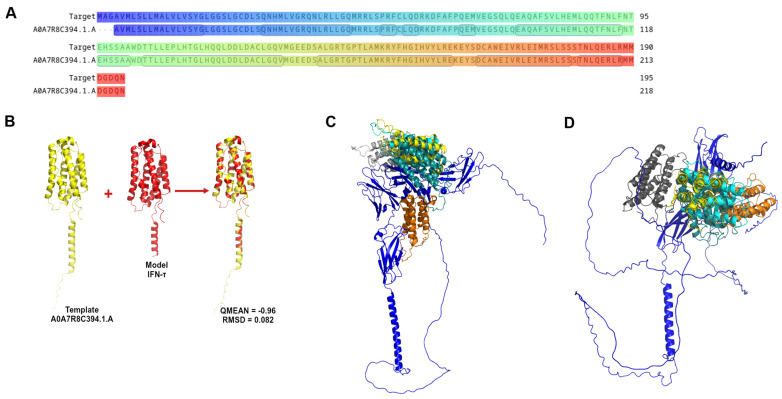
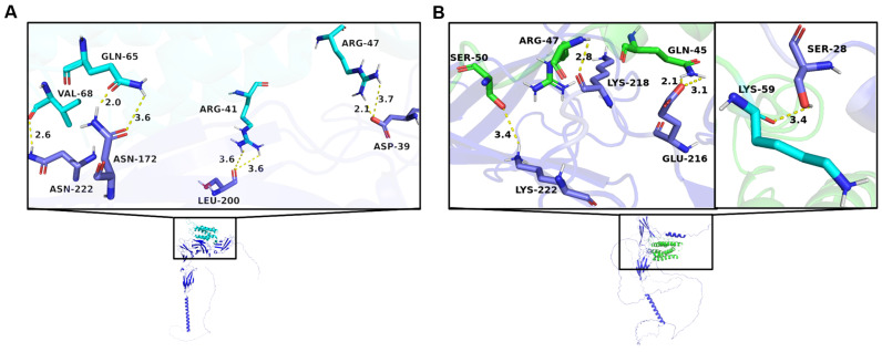
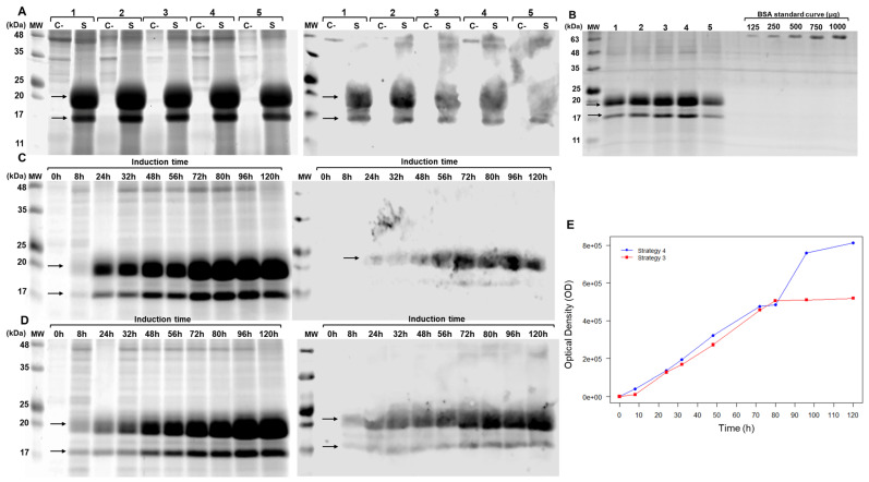
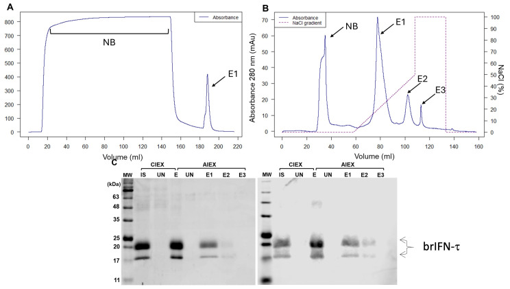
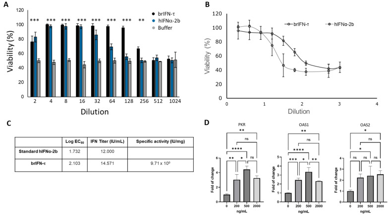
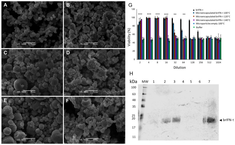
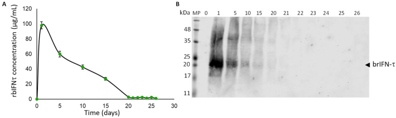
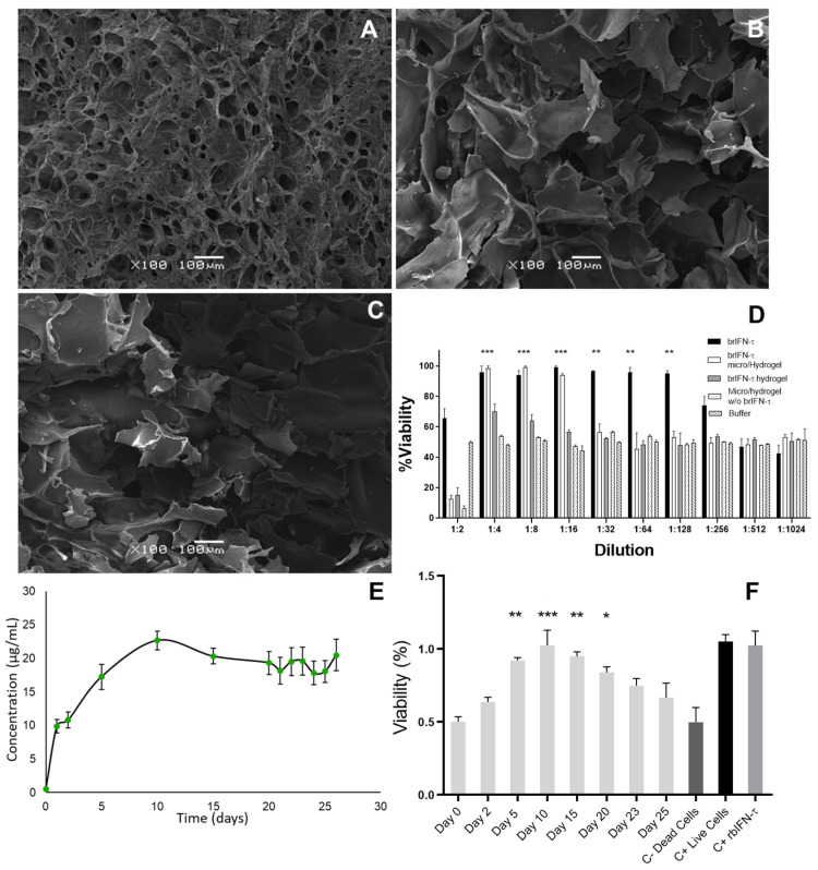
Early embryonic loss is a major cause of reproductive inefficiency in cattle, primarily due to premature luteolysis. Interferon tau (IFN-τ), secreted by the trophoblast, plays a critical role in maternal recognition of pregnancy by maintaining corpus luteum function. However, its practical application has been limited by its rapid degradation and short half-life in vivo. Here, we developed a novel formulation of recombinant bovine IFN-τ, combining chitosan-based microencapsulation with starch-chitosan hydrogel delivery, enabling sustained intrauterine release. This dual-delivery strategy offers a significant improvement over conventional IFN-τ administration methods that rely on repeated intrauterine infusions of soluble protein. The rbIFN-τ was expressed in Pichia pastoris, purified to 90.1% homogeneity, and structurally validated via homology modeling and molecular docking, confirming its interaction with type I interferon receptors. The encapsulated formulation retained antiviral activity, stimulated transcription of interferon-stimulated genes (PKR, OAS1, OAS2), and showed sustained release in vitro for up to 26 days. In vivo evaluation demonstrated safety and biological efficacy, with treated cattle showing inhibited luteolysis, sustained serum progesterone levels, and preserved corpus luteum integrity. This formulation represents a promising biotechnological approach to improve reproductive efficiency through a long-acting, species-specific IFN-τ delivery system.

Keywords: interferon tau; luteolysis modulation; microencapsulation.


Conflict of interest statement

The authors declare no conflicts of interest.

Figures

Figure 1
Figure 1
Graphic representation of IFN-τ sequence and homology modelling. (A) Alignment of the coiled IFN-τ (target: G3MZ51·G3MZ51_BOVIN from UniProt) and the Template A0A7R8C394.1.A. (B) The best model obtained from the Homology Modelling and Docking Experiment. (C) Represents the best poses of α/β-Receptor-1 (blue) with interferon tau. (D) Represents the best poses of α/β-Receptor-2 (blue) with IFN-τ.
Figure 2
Figure 2
Interaction between IFN-τ and receptors. (A) Graphical representation of the main interactions between IFN-τ and α/β-Receptor-1. (B) Graphical representation of the main interactions between IFN-τ and α/β-Receptor-2. The amino acids of α/β-Receptor-1 and 2 are shown in dark blue.
Figure 3
Figure 3
Evaluation of brIFN-τ expression in Pichia pastoris. (A) SDS-PAGE (right) and Western blot (left) of the Pichia pastoris culture supernatant collected 72 h post-induction. Samples are labeled as follows: 1: strategy 1, 2: strategy 2, 3: strategy 3, 4: strategy 4, 5: strategy 5, C-: negative control, S: sample. The arrows indicate the bands corresponding to recombinant bovine IFN-τ. (B) SDS-PAGE gel was used to quantify brIFN-τ from the Pichia pastoris culture supernatant, which was collected 72 h post-induction. Lanes 1–5 represent culture supernatants from strategies 1–5. A BSA standard curve (μg) was used: 125, 250, 500, 750, and 1000 μg. MW: molecular weight markers (kDa). (C) Increasing the culture incubation time to 120 h in shaken flasks. This section includes an SDS-PAGE (right) and Western blot (left) of the culture supernatant using induction strategy 3 at various time points (0–120 h). (D) SDS-PAGE (right) and Western blot (left) using strategy 4 at various time points (0–120 h). (E) brIFN-τ expression curve plotted as a function of time and optical density (OD) for strategies 3 and 4. Original figures can be found in Supplementary Materials.
Figure 4
Figure 4
Evaluation of brIFN-τ purification. (A) Chromatogram from cationic exchange. (B) Chromatogram from anionic exchange. MW: molecular weight, CIEX: cation exchange chromatography, AIEX: anion exchange chromatography, IS: initial sample, UN: unbound proteins to the column, E1: first elution, E2: second elution, E3: third elution fraction. The NaCl gradient used to elute the protein fractions is represented in the graph by a dashed line. (C) SDS-PAGE gel (right) and Western blot (left) of fractions obtained from cationic exchange (S) and anionic exchange (Q) purification. Original figures can be found in Supplementary Materials.
Figure 5
Figure 5
Evaluation of the antiviral activity of brIFN-τ. (A) Percentage of cell viability in inhibiting the cytopathic effect of the Mengo virus in MDBK cells. (B) Antiviral activity of brIFN-τ, with IFNα-2b used as a standard for comparison. (C) Titers of brIFN-τ and specific activity of the standard. (D) As determined by qPCR, relative expression levels of PKR, OAS1, and OAS2. Data were normalized using the Pfaffl method [59,60] with β-actin as the housekeeping gene. Data were statistically analyzed using one-way analysis of variance and Dunnett’s post-test (**** p < 0.0001 *** p < 0.001, ** p < 0.01, * p < 0.05) (ns: p > 0.05).
Figure 6
Figure 6
Microparticle characterization, viability and brIFN-t release. Scanning electron microscope (SEM) photomicrographs of microparticles generated at different temperatures are presented (AF). The empty control particles, which do not contain brIFN-t, were produced at temperatures of 100 °C (A), 120 °C (C), and 140 °C (E). In contrast, brIFN-t associated chitosan particles were generated at the same temperatures: 100 °C (B), 120 °C (D), and 140 °C (F). The viability percentage was assessed by measuring the inhibition of the cytopathic effect of the Mengo virus in MDBK cells (G). A Western blot analysis of microparticle release samples was conducted at 100 °C, 120 °C, and 140 °C (H). The PPM serves as a molecular weight standard, and lanes 1 to 3 correspond to the release supernatants of particle samples generated at 100 °C, 120 °C, and 140 °C, respectively, after 48 h of release in a 10 mM citrate solution at pH 6.5 and 37 °C. Lanes 4 to 6 represent the release samples of empty microparticles generated at 100 °C, 120 °C, and 140 °C, respectively. Lane 7 shows the brIFN-t used as a control (20 µg). A polyclonal antibody from Santa Cruz (Fermelo S.A, Santiago de Chile, Providencia, Chile) was utilized in the experiment, and the reaction was visualized using the Alexa 680 rabbit anti-IgG secondary antibody (Thermo Fisher Scientific, Waltham, MA, USA) Data were statistically analyzed using one-way analysis of variance and Dunnett’s post-test (*** p < 0.001 and ** p < 0.01). Original figures can be found in Supplementary Materials.
Figure 7
Figure 7
Graphical representation of the daily release of brIFN-τ from microparticles generated at 120 °C. (A) Total protein quantification from the released samples daily in the resuspension buffer (50 mM citrate buffer, pH 6.8 and 38 °C to simulate the uterine environment) was performed using the micro-BCA™ Protein Assay Kit. (B) A Western blot analysis was conducted to determine the release of brIFN-τ from chitosan microparticles. The lanes represent the days supernatant samples containing brIFN-τ were extracted. A polyclonal anti-IFN-τ antibody produced in rabbits (Santa Cruz) was utilized, and the reaction was visualized using a secondary antibody, anti-rabbit IgG Alexa 680. The Li-COR Biosciences Odyssey scanner system, along with Image Studio Lite version 5.2 analysis software, was used for visualization and analysis. Original figures can be found in Supplementary Materials.
Figure 8
Figure 8
Scheme of hydrogel generation.
Figure 9
Figure 9
Hydrogel characterization and brIFN-τ release assay. Photomicrographs were taken using scanning electron microscopy (SEM) of the hydrogel samples. (A) Hydrogel mixed with an equivalent of 2 mg of brIFN-τ microencapsulated. (B) Hydrogel mixed with 2 mg of non-microencapsulated soluble brIFN-τ. (C) Hydrogel mixed with empty microparticles. All microparticles were prepared at 120 °C. The brIFN-τ, both microencapsulated and soluble, was added on day 2 of the hydrogel elution scheme, immediately after adding genipin. (D) Viability percentage was determined by measuring the inhibition of the cytopathic effect of the Mengo virus assay in MDBK cells using samples released 48 h after each sample preparation. (E) A graphical representation illustrates the daily release of hydrogel mixed with brIFN-τ microencapsulated over a period of 26 days. Total protein quantification was achieved using the Micro BCA™ Protein Assay Kit. The x-axis corresponds to the day of sampling. (F) This section represents the antiviral effect of brIFN-τ in MDBK cell cultures using ½ dilutions. The samples of hydrogel containing brIFN-τ microencapsulated were utilized for this assay. Results were compared to a negative control of dead cells. Data were statistically analyzed using one-way analysis of variance and Dunnett’s post-test (*** p < 0.001, ** p < 0.01, * p < 0.05).
Figure 10
Figure 10
Drug safety evaluation in the ovine model. (A) Schema of the ovine assay. (B) External evaluation of the genitalia in the study ewes at time 0. Edema and mucosal secretion (yellow arrow). (C) Vulvar edema and erythematous mucosa are due to estrus. (D) Evolution of the rectal temperature in ewes treated with brIFN-τ microencapsulated in hydrogel (brIFN-τ micro/hydrogel) v/s the control group (micro/hydrogel w/o brIFN-τ). (E) Variation in the vulvar temperature in ewes was treated in the same way. Results were compared concerning the negative control of dead cells. No significant differences were registered between the groups.
Figure 11
Figure 11
Anti-luteolytic activity and serum progesterone levels after brIFN-τ delivery via hydrogel in cattle. (A) Scheme of progesterone assay in cows. (B) Transrectal ultrasound evaluation of cows from groups 1, which used microparticles/hydrogel without brIFN-τ, 2 (brIFN-τ microencapsulated with hydrogel), and 3 (non-encapsulated brIFN-τ plus, hydrogel). Visible implant in uterine horn (red arrow); uterine horn (green circle); anechoic center of the uterine horn, indicating the absence of solid contents (green arrow). Ultrasonograph Mindray®, DP50-Vet, linear probe 8.5 MHz. (C) Mold used to generate semi-solid matrix (hydrogel) and hydrogel examples. Sample of implant length (4 cm) (0.3 cm diameter). (D) Progesterone (P4) levels in the three groups: control, brIFN-τ micro/hydrogel, and brIFN-τ hydrogel (nonencapsulated). The levels of P4 on day 19 stand out, with significant differences between brIFN-τ micro/hydrogel concerning the control group. Data were statistically analyzed using one-way analysis of variance and Dunnett’s post-test (*** p < 0.001 and ** p < 0.01).

Similar articles

References

    1. Ealy A.D. Pregnancy Losses in Livestock: An Overview of the Physiology and Endocrinology Symposium for the 2020 Asas-Csas-Wsasas Virtual Meeting. J. Anim. Sci. 2020;98:skaa277. doi: 10.1093/jas/skaa277. - DOI - PMC - PubMed
    1. Sigdel A., Bisinotto R.S., Peñagaricano F. Genes and Pathways Associated with Pregnancy Loss in Dairy Cattle. Sci. Rep. 2021;11:13329. doi: 10.1038/s41598-021-92525-0. - DOI - PMC - PubMed
    1. Williams G.W., Anderson D.P. Choices Magazine 1 The Latin American Livestock Industry: Growth and Challenges. 2020. [(accessed on 1 January 2020)]. Available online: http://www.choicesmagazine.org/choices-magazine/submitted-articles/the-l....
    1. Gädicke P., Monti G. Factors Related to the Level of Occurrence of Bovine Abortion in Chilean Dairy Herds. Prev. Vet. Med. 2013;110:183–189. doi: 10.1016/j.prevetmed.2012.11.022. - DOI - PubMed
    1. Gädicke L’Huissier P., Chihuailaf R., Letelier R., Allende R., Ruiz A., Junod T., Gädicke L’Huissier P., Chihuailaf R., Letelier R., Allende R., et al. Monitoring Different Causal Patterns of Bovine Abortion Syndrome. Vet. México OA. 2022;9:e652. doi: 10.22201/fmvz.24486760e.2022.652. - DOI

MeSH terms

LinkOut - more resources