Review
. 2025 Jul 17;15(7):1036.
doi: 10.3390/biom15071036.

The Intricate Process of Calcification in Granuloma Formation and the Complications Following M. tuberculosis Infection

Affiliations
Review

The Intricate Process of Calcification in Granuloma Formation and the Complications Following M. tuberculosis Infection

Nickolas Yedgarian et al. Biomolecules. .

Abstract

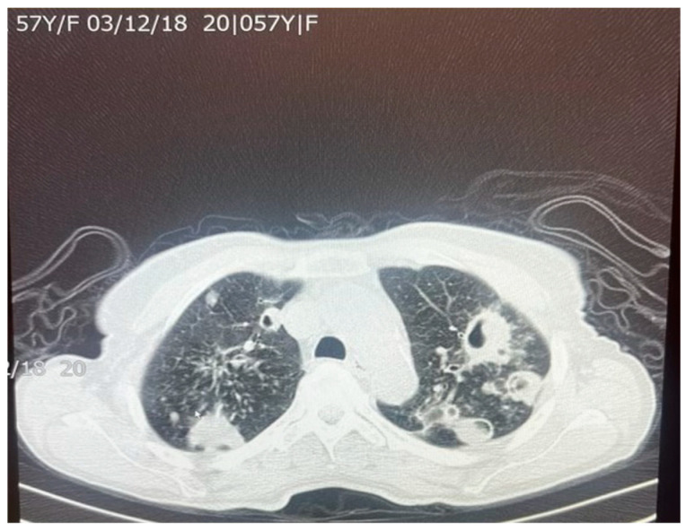
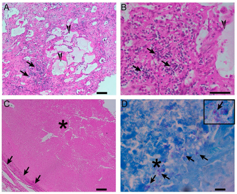
Mycobacterium tuberculosis-an acid-fast staining bacterium-is a serious global health challenge that can have both short-term and long-term complications. Although the immune response helps trap the infection, it can also cause necrosis and calcification, leading to lung tissue damage. Calcification is a known outcome of chronic granuloma evolution in TB. Multiple pathways contribute to fibrosis and calcification; some examples are IL-1β, TGF-β, and TNF-α. Current antifibrotic drugs, such as nintedanib and pirfenidone, are effective but may increase the risk of latent tuberculosis reactivation in certain patients. Experimental therapies such as artemisinin derivatives have shown promise in preclinical TB fibrosis models, while cell-based therapies like bone marrow-derived mononuclear cells are also under early investigation for dual antifibrotic and immunomodulatory effects. This literature review will explore recent studies on the pathogenesis of M. tuberculosis, the mechanisms underlying calcification in granuloma formation, and subsequent complications of the disease process.

Keywords: Mycobacterium tuberculosis; calcification; fibrosis; granuloma.


Conflict of interest statement

The authors declare no conflicts of interest.

Figures

Figure 4
Figure 4
A histological image of a necrotizing granuloma from a pleural biopsy sample of a patient with TB (51 years old /male). The sample is stained with hematoxylin and eosin (H&E), highlighting the central area of caseous necrosis surrounded by epithelioid macrophages, multinucleated giant cells, and a rim of lymphocytes (10× magnification). This structure is characteristic of granulomatous inflammation seen in TB.
Figure 1
Figure 1
(Created in BioRender). Progression of Mycobacterium tuberculosis infection from granuloma formation to scar formation. Infection: Initial infection with Mycobacterium tuberculosis. Acute: CD4+ T helper cells initiate granuloma formation by stimulating TNF-α. Chronic: Chronic inflammation and reactive oxygen species (ROS) release nitric oxide and free radicals, stimulating IL-12 and IFN-γ, leading to bacterial dormancy, collagen formation, and fibrosis. Prolonged Chronic: Prolonged inflammation results in calcified scar formation and fibrosis, encapsulating granuloma. Figure 1 was created with Biorender.com (accessed on 12 April 2025).
Figure 2
Figure 2
Chest X-ray of a 29-year-old male with active TB, showing bilateral upper lobe opacities and cavitation, consistent with advanced pulmonary TB. These findings reflect extensive lung parenchymal damage due to the infection.
Figure 3
Figure 3
Acid-fast bacillus (AFB) staining of a sputum smear from the same patient referenced in Figure 2, demonstrating numerous acid-fast bacilli (red rods) against a blue background (100× magnification).
Figure 5
Figure 5
An axial chest CT scan of a 57-year-old female patient with pulmonary TB, showing bilateral pulmonary cavities and multiple nodules. The cavities are seen predominantly in the upper lobes, with irregular thickened walls, suggestive of active TB. The nodular lesions surrounding the cavities represent granulomatous inflammation, which is characteristic of TB infection. These findings are typical of post-primary TB in adults.
Figure 6
Figure 6
An axial chest CT scan of the same patient in Figure 5, demonstrating advanced nodular and cavitary changes in both lungs, consistent with pulmonary TB. The bilateral upper lobe cavities are clearly visible with associated nodules. The distribution of the lesions, with upper lobe involvement and cavitation, is indicative of reactivation of TB. Additionally, areas of tree-in-bud appearance suggest bronchogenic spread of the infection, which is common in TB patients.
Figure 7
Figure 7
Different types of granulomas, including those comprising diffused immune cell aggregates and mild necrosis (A,B) or fibro-necrotic and caseating nodules (C), were noted on the TB lungs (40× magnification). The former type of granulomas were interspersed with pneumatic fluid accumulation in the lung parenchyma, which was surrounded by mononuclear and polymorphonuclear cells, resembling, respectively, the lymphocytes and neutrophils (A,B). In fibrotic nodules (C), the cellular debris was encapsulated by a fibrotic layer, composed of epithelioid histiocytes. At the core of these lesions, numerous M. tuberculosis-loaded cellular debris were noted ((D) and inset). These observations were consistent with and supported by our previous reports on TB lung granulomas in patients [37,38]. Arrows in panel (A,B) indicate diffused immune cell aggregates. Triangles in panel (A,B) denote mild necrosis. Arrows in panel (C) indicate fibrotic regions. Asteriks in panel (C) illustrate that cellular debris was encapsulated by a fibrotic layer, composed of epithelioid histiocytes. In panel (D), asteriks tllustrate the core of the debris lesions, and arrows denote lesions containing numerous M. tuberculosis-loaded cellular debris.

Similar articles

  • Short-Term Memory Impairment.
    Cascella M, Al Khalili Y. Cascella M, et al. 2024 Jun 8. In: StatPearls [Internet]. Treasure Island (FL): StatPearls Publishing; 2025 Jan–. 2024 Jun 8. In: StatPearls [Internet]. Treasure Island (FL): StatPearls Publishing; 2025 Jan–. PMID: 31424720 Free Books & Documents.
  • Management of urinary stones by experts in stone disease (ESD 2025).
    Papatsoris A, Geavlete B, Radavoi GD, Alameedee M, Almusafer M, Ather MH, Budia A, Cumpanas AA, Kiremi MC, Dellis A, Elhowairis M, Galán-Llopis JA, Geavlete P, Guimerà Garcia J, Isern B, Jinga V, Lopez JM, Mainez JA, Mitsogiannis I, Mora Christian J, Moussa M, Multescu R, Oguz Acar Y, Petkova K, Piñero A, Popov E, Ramos Cebrian M, Rascu S, Siener R, Sountoulides P, Stamatelou K, Syed J, Trinchieri A. Papatsoris A, et al. Arch Ital Urol Androl. 2025 Jun 30;97(2):14085. doi: 10.4081/aiua.2025.14085. Epub 2025 Jun 30. Arch Ital Urol Androl. 2025. PMID: 40583613 Review.
  • The Black Book of Psychotropic Dosing and Monitoring.
    DeBattista C, Schatzberg AF. DeBattista C, et al. Psychopharmacol Bull. 2024 Jul 8;54(3):8-59. Psychopharmacol Bull. 2024. PMID: 38993656 Free PMC article. Review.
  • Systemic pharmacological treatments for chronic plaque psoriasis: a network meta-analysis.
    Sbidian E, Chaimani A, Garcia-Doval I, Do G, Hua C, Mazaud C, Droitcourt C, Hughes C, Ingram JR, Naldi L, Chosidow O, Le Cleach L. Sbidian E, et al. Cochrane Database Syst Rev. 2017 Dec 22;12(12):CD011535. doi: 10.1002/14651858.CD011535.pub2. Cochrane Database Syst Rev. 2017. Update in: Cochrane Database Syst Rev. 2020 Jan 9;1:CD011535. doi: 10.1002/14651858.CD011535.pub3. PMID: 29271481 Free PMC article. Updated.
  • Sexual Harassment and Prevention Training.
    Cedeno R, Bohlen J. Cedeno R, et al. 2024 Mar 29. In: StatPearls [Internet]. Treasure Island (FL): StatPearls Publishing; 2025 Jan–. 2024 Mar 29. In: StatPearls [Internet]. Treasure Island (FL): StatPearls Publishing; 2025 Jan–. PMID: 36508513 Free Books & Documents.

References

    1. Wells W.F., Ratcliffe H.L., Crumb C. On the mechanics of droplet nuclei infection: ii. quantitative experimental air-borne tuberculosis in rabbits1. Am. J. Epidemiol. 1948;47:11–28. doi: 10.1093/oxfordjournals.aje.a119179. - DOI - PubMed
    1. Teitelbaum R., Schubert W., Gunther L., Kress Y., Macaluso F., Pollard J.W., McMurray D.N., Bloom B.R. The M Cell as a Portal of Entry to the Lung for the Bacterial Pathogen Mycobacterium tuberculosis. Immunity. 1999;10:641–650. doi: 10.1016/S1074-7613(00)80063-1. - DOI - PubMed
    1. Flynn J.L., Chan J. Immunology of tuberculosis. Annu. Rev. Immunol. 2001;19:93–129. doi: 10.1146/annurev.immunol.19.1.93. - DOI - PubMed
    1. Ravimohan S., Kornfeld H., Weissman D., Bisson G.P. Tuberculosis and lung damage: From epidemiology to pathophysiology. Eur. Respir. Rev. 2018;27:170077. doi: 10.1183/16000617.0077-2017. - DOI - PMC - PubMed
    1. Guirado E., Schlesinger L.S. Modeling the Mycobacterium tuberculosis Granuloma—The Critical Battlefield in Host Immunity and Disease. Front. Immunol. 2013;4:98. doi: 10.3389/fimmu.2013.00098. - DOI - PMC - PubMed

LinkOut - more resources