. 2025 Jun 20;14(7):628.
doi: 10.3390/antibiotics14070628.

Effect of Pefloxacin on Clostridioides difficile R20291 Persister Cells Formation

Affiliations

Effect of Pefloxacin on Clostridioides difficile R20291 Persister Cells Formation

Camila Queraltó et al. Antibiotics (Basel). .

Abstract

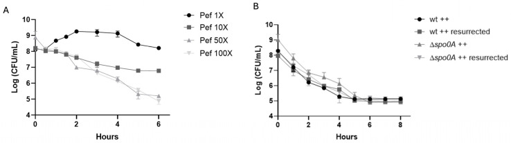
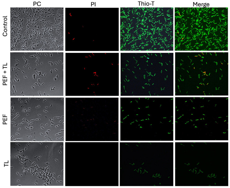
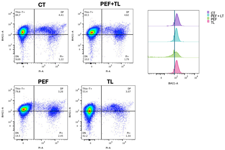
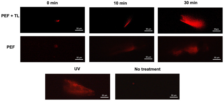
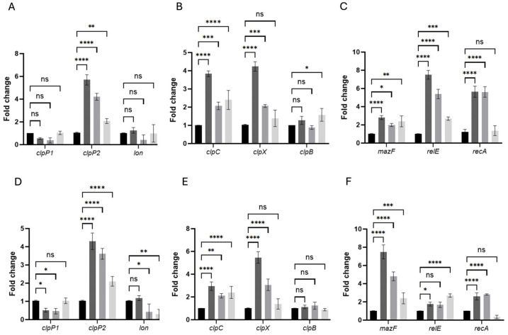
Clostridioides difficile is a Gram-positive bacterium recognized for its ability to produce toxins and form spores. It is mainly accountable for the majority of instances of antibiotic-related diarrhea. Background. Bacterial persister represent a minor fraction of the population that shows temporary tolerance to bactericidal agents, and they pose considerable medical issues because of their link to the rise of antibiotic resistance and challenging chronic or recurrent infections. Our previous research has shown a persister-like phenotype associated with treatments that include pefloxacin. Nonetheless, the mechanism is still mostly unclear, mainly because of the difficulty in isolating this small group of cells. Objectives. To enhance the understanding of C. difficile persister cells, we made an enrichment and characterization of these cells from bacterial cultures during the exponential phase under pefloxacin treatment and lysis treatment. Results. We demonstrate the appearance of cells with lower metabolism and DNA damage. Furthermore, we noted the participation of toxin-antitoxin systems and Clp proteases in the generation of persister cells. Conclusions. This work demonstrates the formation of C. difficile persister cells triggered by a lethal concentration of pefloxacin.

Keywords: Clostridioides difficile; pefloxacin; persister cells.


Conflict of interest statement

The authors declare no conflicts of interest.

Figures

Figure 1
Figure 1
Pefloxacin-induced persistence in C. difficile R20291. (A) Time–kill kinetics of exponential-phase cultures treated with pefloxacin at 1×, 10×, 50×, and 100× the determined MIC (1.53 µg/mL). High-dose treatments (50× MIC only) display a characteristic biphasic killing curve: an initial rapid decline in viable counts followed by a persistent plateau (n = 3; mean ± SD). (B) Rechallenge of survivors from the 50× MIC treatment and comparison of wild-type (wt) versus non-sporulating Δspo0A mutant under identical conditions. Cultures were washed, resuspended in BHIS++ (supplemented with taurocholate, fructose, and glucose), and exposed again to 50× MIC pefloxacin (76.5 µg/mL). The reproduced biphasic kill profile and unchanged MIC confirm a reversible, non-sporulating persister phenotype (n = 3; mean ± SD).
Figure 2
Figure 2
Lysis-enriched persister cell survival in C. difficile R20291. (A) Basal persister frequency following lysis without antibiotic exposure. Wild-type (black bars) and Δspo0A (gray bars) cultures were treated with lysis buffer alone, yielding a low, time-independent survival of ~1.8% (n = 3; mean ± SD), consistent with stochastic (type I) persister formation. (B) Antibiotic-induced persister enrichment. Cultures pre-treated with 50× MIC pefloxacin (76.5 µg/mL) for 30 min, 1, 2, and 4 h were washed, subjected to the same 15 min lysis, and plated for CFU. Survival increased in a time-dependent manner, peaking at ~10% after 2 h and remaining stable through 4 h (n = 3; mean ± SD). No significant difference was observed between wt and Δspo0A at any time point (ns), indicating that persister enrichment is independent of Spo0A-mediated sporulation. (** p < 0.005).
Figure 3
Figure 3
Fluorescence assessment of metabolic activity and membrane integrity in persister-enriched C. difficile R20291. Wild-type R20291 cultures in exponential phase were subjected to one of four conditions: no treatment (Control), lysis buffer only (TL), pefloxacin at 50× MIC only (PEF), or pefloxacin followed by lysis (PEF + TL). Cells were (left) imaged by phase-contrast (CF), (middle) stained with PI (red) to reveal membrane permeability, and (right) stained with Thio-T (green) to report RNA metabolic activity (n = 3).
Figure 4
Figure 4
Flow cytometric profiling of C. difficile persister induction and enrichment. (Left) Representative dual-parameter dot plots of exponential-phase cultures: untreated control (CT), pefloxacin alone (PEF), lysis treatment alone (TL), and combined pefloxacin + lysis (PEF + TL). Cells were stained with Thio-T (metabolic activity) and PI (membrane integrity). Quadrants delineate Thio-T+ only (upper left), PI+ only (lower right), double-negative (DN; lower left) and double-positive (DP; upper right) subpopulations, with percentages indicated. (Right) Overlaid histograms of Thio-T fluorescence for each condition. The dashed vertical line marks the threshold separating Thio-T and Thio-T+ signals.
Figure 5
Figure 5
DNA degradation in pefloxacin-induced C. difficile enriched persister cells. Comet assay performed on persister cells enriched by lysis treatment and previously exposed to pefloxacin 50× (top panel) and cells exposed to pefloxacin 50× without lysis treatment (middle panel) for 0, 15 and 30 min. Untreated cells were used as negative control and cells treated with UV radiation for 10 min were used as positive control (lower panel) (n = 3). Images of 5 fields were taken and the most representative was selected.
Figure 6
Figure 6
Persistence-associated genes are rapidly induced in pefloxacin-enriched C. difficile persister cells. RNA was extracted at 10 min (AC) and 30 min (DF) after treatment with 50× MIC pefloxacin following lysis enrichment. Transcript levels of the peptidase genes clpP1, clpP2 and the lon protease (A,D), the chaperone genes clpC, clpX and clpB (B,E), and the toxin–antitoxin components mazF, relE as well as the SOS regulator recA (C,F) were measured by RT-qPCR and normalized to 16S rRNA. Data are presented as mean ± SD of three biological replicates (n = 3), with statistical significance relative to untreated controls indicated as * p < 0.01, ** p < 0.005, *** p < 0.001, **** p < 0.0001, and ns (not significant). Black bars, untreated control (CT); dark-gray bars, pefloxacin + lysis (PEF + TL); medium-gray bars, pefloxacin alone (PEF); light-gray bars, lysis alone (TL).

Similar articles

References

    1. Cole S.A., Stahl T.J. Persistent and Recurrent Clostridium difficile Colitis. Clin. Colon Rectal Surg. 2015;28:065–069. doi: 10.1055/s-0035-1547333. - DOI - PMC - PubMed
    1. Deneve C., Janoir C., Poilane I., Fantinato C., Collignon A. New trends in Clostridium difficile virulence and pathogenesis. Int. J. Antimicrob. Agents. 2009;33:S24–S28. doi: 10.1016/S0924-8579(09)70012-3. - DOI - PubMed
    1. Karen C.C., John G.B. Biology of Clostridium difficile: Implications for epidemiology and diagnosis. Annu. Rev. Microbiol. 2011;65:501–521. doi: 10.1146/annurev-micro-090110-102824. - DOI - PubMed
    1. Boven A., Vlieghe E., Engstrand L., Andersson F.L., Callens S., Simin J., Brusselaers N. Clostridioides difficile infection-associated cause-specific and all-cause mortality: A population-based cohort study. Clin. Microbiol. Infect. 2023;29:1424–1430. doi: 10.1016/j.cmi.2023.07.008. - DOI - PubMed
    1. Di Bella S., Sanson G., Monticelli J., Zerbato V., Principe L., Giuffrè M., Luzzati R. Clostridioides difficile infection: History, epidemiology, risk factors, prevention, clinical manifestations, treatment, and future options. Clin. Microbiol. Rev. 2024;37:e0013523. doi: 10.1128/cmr.00135-23. - DOI - PMC - PubMed

Grants and funding

LinkOut - more resources