. 2025 Jun 23;14(7):640.
doi: 10.3390/antibiotics14070640.

Dual Redox Targeting by Pyrroloformamide A and Silver Ions Enhances Antibacterial and Anti-Biofilm Activity Against Carbapenem-Resistant Klebsiella pneumoniae

Affiliations

Dual Redox Targeting by Pyrroloformamide A and Silver Ions Enhances Antibacterial and Anti-Biofilm Activity Against Carbapenem-Resistant Klebsiella pneumoniae

Enhe Bai et al. Antibiotics (Basel). .

Abstract

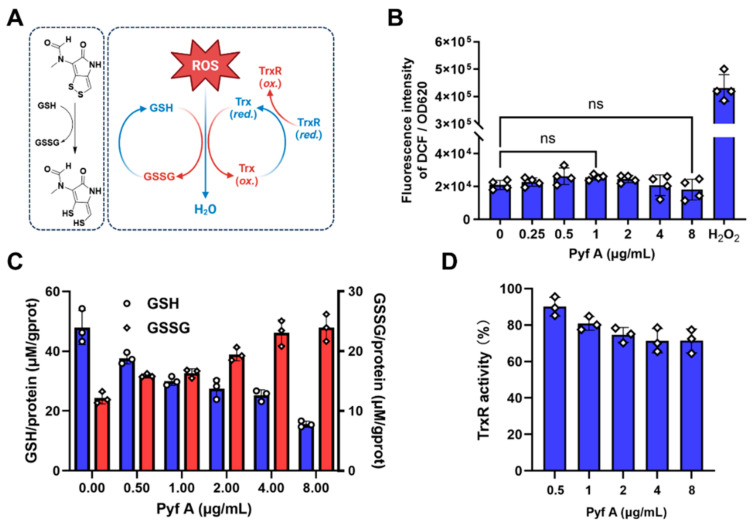
Background: Dithiolopyrrolones (DTPs), such as holomycin and thiolutin, exhibit potent antibacterial activities. DTPs contain a disulfide within a unique bicyclic scaffold, which may chelate metal ions and disrupt metal-dependent cellular processes once the disulfide is reductively transformed to thiols. However, the contribution of the intrinsic redox mechanism of DTPs to their antibacterial activity remains unclear. Herein we used pyrroloformamide (Pyf) A, a DTP with a unique formyl substituent, as a prototype to study the antibacterial potential and mechanism against ESKAPE pathogens, in particular carbapenem-resistant Klebsiella pneumoniae (CRKP). Methods: The antibacterial and anti-biofilm activities of Pyf A were mainly assessed against clinical CRKP isolates. Propidium iodide staining, scanning electron microscopy, glutathione (GSH) quantification, and reactive oxygen species (ROS) analysis were utilized to infer its anti-CRKP mechanism. The synergistic antibacterial effects of Pyf A and AgNO3 were evaluated through checkerboard and time-kill assays, as well as in vivo murine wound and catheter biofilm infection models. Results: Pyf A exhibited broad-spectrum antibacterial activity against ESKAPE pathogens with minimum inhibitory concentrations ranging from 0.25 to 4 μg/mL. It also showed potent anti-biofilm effects against CRKP. Pyf A disrupted the cell membranes of CRKP and markedly depleted intracellular GSH without triggering ROS accumulation. Pyf A and AgNO3 showed synergistic anti-CRKP activities in vitro and in vivo, by disrupting both GSH- and thioredoxin-mediated redox homeostasis. Conclusions: Pyf A acts as a GSH-depleting agent and, when combined with AgNO3, achieves dual-targeted disruption of bacterial thiol redox systems. This dual-targeting strategy enhances antibacterial efficacy of Pyf A and represents a promising therapeutic approach to combat CRKP infections.

Keywords: AgNO3; carbapenem-resistant Klebsiella pneumoniae; glutathione; pyrroloformamide A; redox homeostasis; thioredoxin.


Conflict of interest statement

The authors declare no conflicts of interest.

Figures

Figure 1
Figure 1
The structure and mode of actions of dithiolopyrrolones and related natural products. (A) Representative dithiolopyrrolones and natural products containing redox-active sulfur atoms. (B) The known mode of action of the prototypical holomycin: it followed reductive activation and formation of a Zn+ containing metallocomplex to inhibit zinc-dependent metalloproteins and disrupt intracellular metal homeostasis.
Figure 2
Figure 2
Antibacterial activity of Pyf A against clinical multidrug-resistant pathogens. (A) Minimum inhibitory concentrations (MICs) of Pyf A and five conventional antibiotics against ‘ESKAPE’ clinical isolates. (B) Time-kill curves of Pyf A against KP113 at concentrations of 2, 4, 8, and 16 μg/mL. (C) Time-kill curves of Pyf A against MDR SA116 at concentrations of 0.25, 0.5, 1, and 2 μg/mL.
Figure 3
Figure 3
Effects of Pyf A on biofilm formation in KP113. (A) Inhibition of biofilm formation by Pyf A at various concentrations. Biofilm biomass was quantified by crystal violet staining (OD540), and bacterial load in the supernatant was measured by CFU counting. (B) Disruption of pre-formed mature biofilms by Pyf A. After 48 h biofilm formation, biofilms were treated with Pyf A for 12 h, followed by biomass and bacterial load quantification as in (A) (n = 6). (C) Bright-field microscopy images showing structural changes in biofilms following treatment with increasing concentrations of Pyf A (0, 2, 8, and 16 μg/mL). Scale bar: 500 μm. Data in (A) and (B) represent mean ± SD (n = 6).
Figure 4
Figure 4
Pyf A disrupts the membrane integrity of K. pneumoniae in a dose- and time-dependent manner. (A) KP113 was treated with increasing concentrations of Pyf A (2, 4, and 8 μg/mL) for 4 h. Membrane damage was assessed by propidium iodide (PI) staining and scanning electron microscopy (SEM). (B) Quantification of PI fluorescence intensity after 4 h treatment with Pyf A, normalized to OD600. Data represent mean ± SD (n = 3), dots represent individual data points. (C) Time-course of PI fluorescence intensity in K. pneumoniae treated with Pyf A at 2 and 4 μg/mL.
Figure 5
Figure 5
Pyf A depletes glutathione without inducing ROS accumulation and exhibits partial TrxR inhibitory activity. (A) Schematic of the cellular antioxidant systems and their interplay in scavenging ROS. Pyf A is proposed to disrupt the glutathione (GSH/GSSG) systems, potentially leading to oxidative imbalance. (B) Measurement of intracellular ROS levels in KP113 after treatment with Pyf A. H2O2 (1 mM) was used as a positive control (n = 4). (C) Quantification of intracellular GSH and GSSG levels in KP113 treated with Pyf A. blue bars represent GSH levels, and red bars represent GSSG levels. Pyf A induced a dose-dependent decrease in GSH and increase in GSSG. (D) Pyf A modestly inhibit TrxR enzymatic activity (n = 3). Data represent mean ± SD; ns, not significant.
Figure 6
Figure 6
Synergistic bactericidal activity of Pyf A and AgNO3 through disruption of redox homeostasis and induction of ROS. (A) Proposed mechanism of ROS generation and redox system disruption by Pyf A and AgNO3. Pyf A perturbs the glutathione (GSH/GSSG) cycle, while AgNO3 inhibits the thioredoxin (Trx/TrxR) system. Their combined action leads to oxidative stress and ROS accumulation. (B) Heat map showing the bacterial viability (CFU/mL) under different concentrations of Pyf A and AgNO3. Darker shades indicate higher CFU counts. (C) Isobologram showing the fractional inhibitory concentration (FIC) indices of Pyf A and AgNO3. Data points below the green line indicate synergistic interaction (FIC index < 0.5). (D) Time-kill curves of KP113 treated with Pyf A, AgNO3, or their combination. The combination led to a rapid and sustained decrease in bacterial load below the detection limit. Data represent mean ± SD (n = 3).
Figure 7
Figure 7
Combination of Pyf A and AgNO3 induces ROS accumulation, redox imbalance, and enhanced antibacterial activity. (A) Intracellular ROS levels measured in KP113 treated with Pyf A (0.5 μg/mL), AgNO3 (0.5 μg/mL), or their combination. H2O2 (1 mM) served as a positive control. (B) Intracellular levels of reduced (GSH) and oxidized (GSSG) glutathione upon indicated treatments. blue bars represent GSH levels, and red represent GSSG. (C) Dose-dependent inhibition of TrxR activity by AgNO3. (D) TrxR activity in bacterial lysates treated with Pyf A (0.5 μg/mL), AgNO3 (0.5 μg/mL), or both. (E) Heatmap showing bacterial viability (CFU/mL) after treatment with various combinations of Pyf A and AgNO3. Lighter shades indicate greater antibacterial activity. Data represent mean ± SD (n = 3); p < 0.01 (**).
Figure 8
Figure 8
The anti-biofilm effects of Pyf A and AgNO3 against KP113. (A) Inhibition of biofilm formation by Pyf A (0.5 μg/mL), AgNO3 (0.5 μg/mL), or their combination. Biofilm biomass was quantified by crystal violet staining (OD450), and bacterial load in the supernatant was measured by CFU counting (n = 6). (B) Disruption of pre-formed mature biofilms by Pyf A (0.5 μg/mL), AgNO3 (0.5 μg/mL), or their combination. After 48 h biofilm formation, biofilms were treated for 12 h, followed by biomass and bacterial load quantification as in (A) (n = 6). (C) Representative fluorescence microscopy images of PI-stained biofilms. Red fluorescence denotes dead or membrane-damaged cells. Scale bar = 200 μm.
Figure 9
Figure 9
Pyf A combined with AgNO3 promotes wound healing and reduces bacterial burden in a murine skin infection model by K. pneumoniae. (A) The schematic form of the wound infection model. Animals were subjected to full-thickness excisional wounds and inoculated with KP113 (1 × 10⁶ CFU/wound, 24 h), followed by topical treatment from day 1 to day 14. Pyf A ointment (0.5 mg/mL), AgNO3 (0.5 mg/mL), combination therapy (Pyf A 0.5 mg/mL + AgNO3 0.5 mg/mL), and imipenem (1 mg/mL). The vehicle control and infected group received blank ointment base. (B) Bacterial burden in infected tissues (log CFU/g) after 7 days of treatment. The combination significantly reduced bacterial load, compared to single-drug and imipenem (n = 5). (C) Representative images of wounds at day 0, 7, and 14 post-treatment, alongside corresponding wound size maps. The combination group showed markedly improved healing and smaller wound areas (n = 3). Data represent mean ± SD; p < 0.01 (**).
Figure 10
Figure 10
Combination of Pyf A and AgNO3 effectively eradicates biofilm-associated catheter infection model of K. pneumoniae. (A) Schematic of catheter-associate biofilm model. The treatment groups included the following: Pyf A (0.5 mg/mL), AgNO3 (0.5 mg/mL), combination therapy (Pyf A 0.5 mg/mL + AgNO3 0.5 mg/mL), and vehicle control (saline). (B) Representative crystal violet-stained images of catheters collected at the endpoint of the experiment. (C) Bacterial load (log10 CFU/mL) on catheters after 14 days. (D) Biofilm biomass quantification: crystal violet-stained catheters from each animal were washed, destained with ethanol, and measured at OD540. Data represent mean ± SD (n = 4), p < 0.01 (**).

Similar articles

References

    1. Qiao W., Wang L., Luo Y., Yang T. Synthetic approaches and therapeutic applications of FDA-approved antibacterial agents: A comprehensive review from 2003 to 2023. Eur. J. Med. Chem. 2025;285:117267–117291. doi: 10.1016/j.ejmech.2025.117267. - DOI - PubMed
    1. Kinch M.S., Patridge E., Plummer M., Hoyer D. An analysis of FDA-approved drugs for infectious disease: Antibacterial agents. Drug Discov. Today. 2014;19:1283–1287. doi: 10.1016/j.drudis.2014.07.005. - DOI - PubMed
    1. Murray C.J.L., Ikuta K.S., Sharara F., Swetschinski L., Robles Aguilar G., Gray A., Han C., Bisignano C., Rao P., Wool E., et al. Global burden of bacterial antimicrobial resistance in 2019: A systematic analysis. Lancet. 2022;399:629–655. doi: 10.1016/S0140-6736(21)02724-0. - DOI - PMC - PubMed
    1. WHO Bacterial Priority Pathogens List, 2024: Bacterial Pathogens of Public Health Importance to Guide Research, Development and Strategies to Prevent and Control Antimicrobial Resistance. 2024. [(accessed on 17 May 2025)]. Available online: https://www.who.int/publications/i/item/9789240093461. - PubMed
    1. Antimicrobial Resistance, Hypervirulent Klebsiella pneumoniae—Global Situation. 2024. [(accessed on 17 May 2025)]. Available online: https://www.who.int/emergencies/disease-outbreak-news/item/2024-DON527.

LinkOut - more resources