

Super-Resolution Contrast-Enhanced Ultrasound Examination Down to the Microvasculature Enables Quantitative Analysis of Liver Lesions: First Results
- PMID: 40724493
- PMCID: PMC12299643
- DOI: 10.3390/life15070991
Super-Resolution Contrast-Enhanced Ultrasound Examination Down to the Microvasculature Enables Quantitative Analysis of Liver Lesions: First Results
Abstract
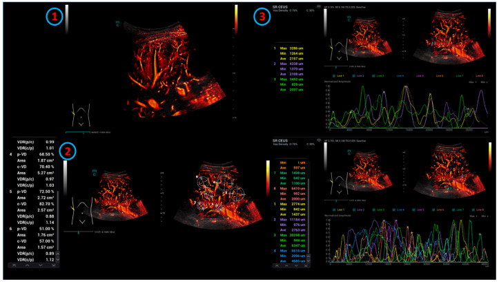
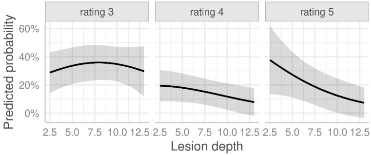
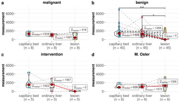
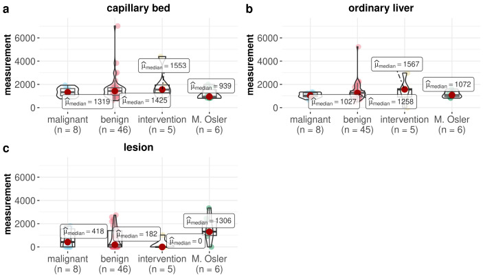
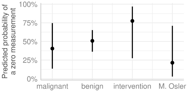
This article investigates the first use of dynamic microvascularization of liver lesions in clinical evaluation using real-time super-resolution contrast-enhanced ultrasound (SR CEUS). A retrospective analysis of SR CEUS examinations of liver lesions was performed. All examinations were conducted using an SC7-1U convex probe after the bolus injection of the ultrasound contrast agent. Digital cine loops were stored for independent evaluation. The evaluation was performed with respect to parallel measuring lines (the diameter corresponded to the capillary density) in the area of the reference lesion, liver tissue, and liver capsule using statistical analysis. In total, 65 patients (female n = 28; male n = 37; average age 57.8 ± 17.2 years) were evaluated. The examined liver lesions were mostly benign masses (n = 46). Mild liver fibrosis (stage F1) was present in most cases (n = 35). The lesions examined were located at an average tissue depth of 6.07 ± 2.47 cm. The highest number of lesion measurements was observed in the malignant lesion group. Significant differences in the measurements were found when comparing benign lesions with the capillary region (p < 0.001) and normal liver tissue (p < 0.01). The use of SR CEUS opens up new possibilities for the quantification of neovascularization, assessment of microvascular changes, and evaluation of the follow-up of intrahepatic interventions.
Keywords: contrast agent; intervention; liver; microvascularization; ultrasonography.
Conflict of interest statement
All authors declare no conflicts of interest.
Figures
Similar articles
-
Contrast-enhanced ultrasound using SonoVue® (sulphur hexafluoride microbubbles) compared with contrast-enhanced computed tomography and contrast-enhanced magnetic resonance imaging for the characterisation of focal liver lesions and detection of liver metastases: a systematic review and cost-effectiveness analysis.Health Technol Assess. 2013 Apr;17(16):1-243. doi: 10.3310/hta17160. Health Technol Assess. 2013. PMID: 23611316 Free PMC article.
-
Contrast-enhanced ultrasound for the diagnosis of hepatocellular carcinoma in adults with chronic liver disease.Cochrane Database Syst Rev. 2022 Sep 2;9(9):CD013483. doi: 10.1002/14651858.CD013483.pub2. Cochrane Database Syst Rev. 2022. PMID: 36053210 Free PMC article.
-
The application of multimodal ultrasound examination in the differential diagnosis of benign and malignant breast lesions of BI-RADS category 4.Front Med (Lausanne). 2025 Jun 9;12:1596100. doi: 10.3389/fmed.2025.1596100. eCollection 2025. Front Med (Lausanne). 2025. PMID: 40552183 Free PMC article.
-
Contrast-enhanced ultrasound-based AI model for multi-classification of focal liver lesions.J Hepatol. 2025 Aug;83(2):426-439. doi: 10.1016/j.jhep.2025.01.011. Epub 2025 Jan 21. J Hepatol. 2025. PMID: 39848548
-
Adefovir dipivoxil and pegylated interferon alfa-2a for the treatment of chronic hepatitis B: a systematic review and economic evaluation.Health Technol Assess. 2006 Aug;10(28):iii-iv, xi-xiv, 1-183. doi: 10.3310/hta10280. Health Technol Assess. 2006. PMID: 16904047
References
-
- Jung E.M., Kaiser U., Herr W., Stroszczynski C., Jung F. Novel high-resolution contrast agent ultrasound techniques HiFR CEUS and SR CEUS in combination with shear wave elastography, fat assessment and viscosity of liver parenchymal changes and tumors. Clin. Hemorheol. Microcirc. 2024;86:263–273. doi: 10.3233/CH-249103. - DOI - PubMed
-
- Pausch A.M., Kammerer S., Weber F., Herr W., Stroszczynski C., Holler E., Edinger M., Wolff D., Weber D., Jung E.M., et al. Parametric Imaging of Contrast-Enhanced Ultrasound (CEUS) for the Evaluation of Acute Gastrointestinal Graft-Versus-Host Disease. Cells. 2021;10:1092. doi: 10.3390/cells10051092. - DOI - PMC - PubMed
LinkOut - more resources
Full Text Sources
Research Materials
