. 2025 Jun 23;15(7):998.
doi: 10.3390/life15070998.

Feasibility of Xenogeneic Mitochondrial Transplantation in Neuronal Systems: An Exploratory Study

Affiliations

Feasibility of Xenogeneic Mitochondrial Transplantation in Neuronal Systems: An Exploratory Study

Eriko Nakamura et al. Life (Basel). .

Abstract

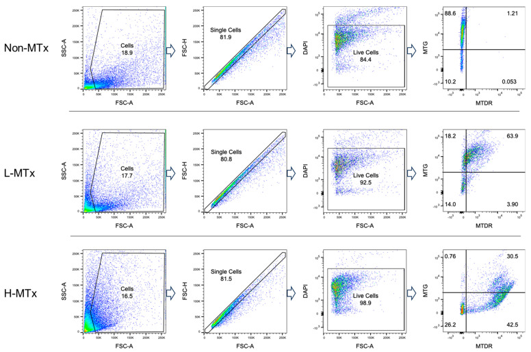
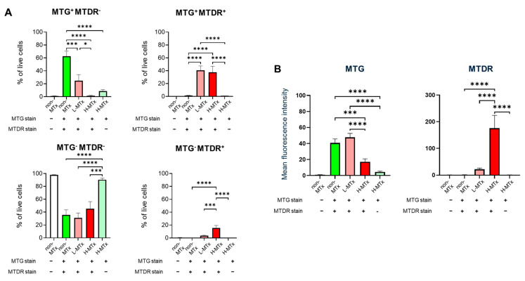
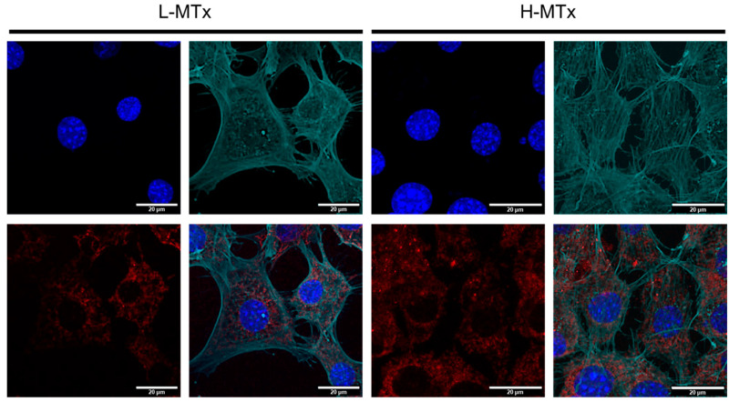
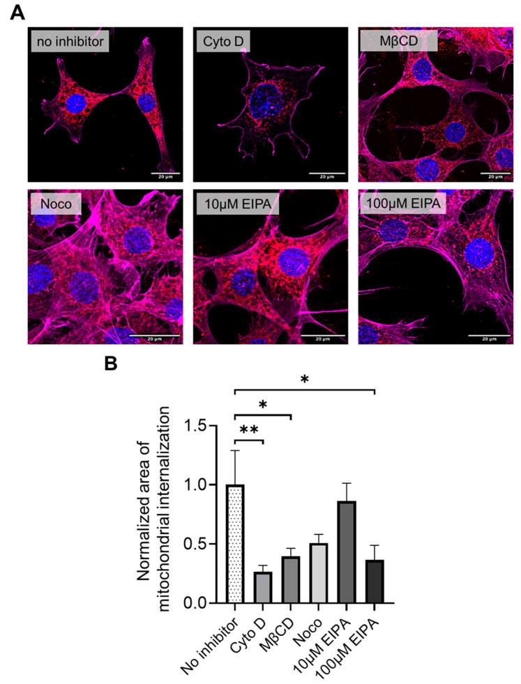
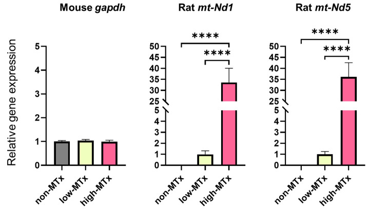
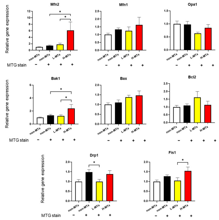
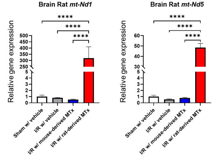
Mitochondrial transplantation (MTx) has emerged as a potential therapeutic approach for diseases associated with mitochondrial dysfunction, yet its scalability and cross-species feasibility remain underexplored. This study aimed to evaluate the dose-dependent uptake and molecular effects of xenogeneic mitochondrial transplantation (xeno-MTx) using rat-derived mitochondria in mouse neuronal systems. HT-22 hippocampal neuronal cells and a murine model of cardiac arrest-induced global cerebral ischemia were used to assess mitochondrial uptake, gene expression, and mitochondrial DNA presence. Donor mitochondria were isolated from rat pectoralis muscle and labeled with MitoTracker dyes. Flow cytometry and confocal microscopy revealed a dose-dependent increase in donor mitochondrial uptake in vitro. Quantitative PCR demonstrated a corresponding increase in rat-specific mitochondrial DNA and upregulation of Mfn2 and Bak1, with no changes in other fusion, fission, or apoptotic genes. Inhibitor studies indicated that mitochondrial internalization may involve actin-dependent macropinocytosis and cholesterol-sensitive endocytic pathways. In vivo, rat mitochondrial DNA was detected in mouse brains post-xeno-MTx, confirming donor mitochondrial delivery to ischemic tissue. These findings support the feasibility of xeno-MTx and its dose-responsive biological effects in neuronal systems while underscoring the need for further research to determine long-term functional outcomes and clinical applicability.

Keywords: cardiac arrest; ischemia–reperfusion; mitochondria; mitochondrial transplantation; neuron.


Conflict of interest statement

The authors declare no conflict of interest.

Figures

Figure 1
Figure 1
Transmission electron microscopy of isolated mitochondria. Representative transmission electron microscopy images showing rat-derived mitochondria with preserved morphology, including intact outer membranes and visible cristae structures. These features are consistent with the structural integrity of the isolated mitochondria. Scale bar = 600 nm.
Figure 2
Figure 2
Representative flow cytometry gating strategy for neuronal cell populations labeled with MitoTracker dyes. HT-22 murine hippocampal neuronal cells were stained with MitoTracker Green (MTG) to label endogenous mitochondria and MitoTracker Deep Red (MTDR) to label exogenous donor mitochondria. The gating illustrates three distinct cell populations: MTG+MTDR (cells containing only endogenous mitochondria), MTG+MTDR+ (cells containing both endogenous and donor mitochondria), and MTGMTDR+ (cells containing primarily donor mitochondria). Dead cells were excluded by DAPI staining, and fluorescence thresholds were defined using unstained controls.
Figure 3
Figure 3
Dose-dependent uptake of donor mitochondria by HT-22 neuronal cells following xenogeneic mitochondrial transplantation. (A) Quantitative analysis of HT-22 cell populations shows a dose-dependent decrease in MTG+MTDR cells and corresponding increases in MTG+MTDR+ and MTGMTDR+ cells following exposure to low-dose (L-MTx: 10 µg/mL) and high-dose (H-MTx: 1 mg/mL) donor mitochondria. (B) Mean fluorescence intensity (MFI) analysis of MTDR and MTG signals. MTDR MFI increased significantly with higher mitochondrial doses, while MTG MFI decreased following xeno-MTx treatment. Fluorescence intensity was used as a relative surrogate of mitochondrial uptake, although overlap between MTG and MTDR signals may limit interpretability. Data represent mean ± SEM (n = 9 per group). * p < 0.05, *** p < 0.001, **** p < 0.0001 between each group.
Figure 4
Figure 4
Confocal microscopy showing dose-dependent uptake of donor mitochondria by HT-22 hippocampal neuronal cells. Representative confocal images of HT-22 cells following exposure to low-dose (L-MTx: 10 µg/mL) and high-dose (H-MTx: 1 mg/mL) rat-derived mitochondria labeled with MitoTracker Deep Red (MTDR; red). The actin cytoskeleton is pseudo-colored green to highlight cell morphology and aid in visualizing mitochondrial localization. Nuclei were counterstained with DAPI (blue). Cells treated with L-MTx showed mild-to-moderate MTDR signal, whereas cells treated with H-MTx exhibited more intense and widespread MTDR fluorescence, consistent with a dose-dependent increase in donor mitochondrial presence. Endogenous mitochondria were not labeled in these experiments to avoid signal overlap with exogenous MTDR. Scale bar = 20 μm.
Figure 5
Figure 5
Pharmacological inhibition of mitochondrial internalization pathways in HT-22 cells during xeno-MTx. (A) Representative fluorescence microscopy images of HT-22 neuronal cells exposed to MTDR-labeled donor mitochondria following pretreatment with pathway-specific inhibitors. Donor mitochondria are visualized in red (MTDR), nuclei in blue (DAPI), and the actin cytoskeleton is pseudo-colored in magenta in this figure (note: pseudo-colored green was used in Figure 4). Reduced MTDR fluorescence was observed after pretreatment with cytochalasin D (actin polymerization inhibitor), methyl-β-cyclodextrin (MβCD; cholesterol-depleting agent), and high-dose EIPA (100 µM; macropinocytosis inhibitor), compared to untreated controls. Pretreatment with nocodazole (microtubule destabilizer) or low-dose EIPA (10 µM) did not markedly affect mitochondrial uptake. (B) Quantitative analysis of cellular MTDR signal intensity confirms significant reductions in mitochondrial uptake following treatment with cytochalasin D, MβCD, and high-dose EIPA. Data represent mean ± SEM (n = 12 per group). * p < 0.05, ** p < 0.01 between each group.
Figure 6
Figure 6
Detection of rat-specific mitochondrial DNA in recipient neuronal cells following in vitro xeno-MTx. Quantitative real-time PCR detected rat mitochondrial genes (mt-Nd1 and mt-Nd5) exclusively in HT-22 neuronal cells treated with donor mitochondria (low dose: 0.1 µg/mL; high dose: 10 µg/mL), but not in untreated controls. A statistically significant positive linear relationship was observed between mitochondrial dose and gene copy number for both mt-Nd1 and mt-Nd5 (linear trend test, **** p < 0.0001). These results are consistent with a dose-dependent increase in the presence of donor mitochondrial DNA in recipient cells. Data are presented as mean ± SEM (n = 6 per group).
Figure 7
Figure 7
Effects of xeno-MTx on expression of mitochondrial dynamics and apoptosis-related genes in neuronal cells in vitro. Quantitative real-time PCR analysis of HT-22 cells treated with low- or high-dose donor mitochondria revealed a dose-dependent increase in the expression of the mitochondrial fusion gene Mfn2 and the pro-apoptotic gene Bak1. No significant dose-dependent changes were observed in other genes related to mitochondrial fission (Dnm1l, Fis1), fusion (Opa1, Mfn1), or apoptosis (Bax, Bcl2). Data represent mean ± SEM (n = 10–13 per group). * p < 0.05 between each group.
Figure 8
Figure 8
Detection of donor mitochondrial DNA following xeno-MTx in an in vivo mouse model of global cerebral ischemia–reperfusion. Quantitative real-time PCR analysis detected rat-specific mitochondrial genes (mt-Nd1 and mt-Nd5) in the brains of mice that received intravenous donor mitochondria after cardiac arrest and resuscitation. Significantly higher levels of rat mitochondrial DNA were observed in xeno-MTx–treated mice compared to sham-operated controls. These results are consistent with successful delivery and retention of donor mitochondria in ischemic brain tissue. Data presented as mean ± SEM (n = 3–6 per group). **** p < 0.0001 between each group.

Similar articles

References

    1. Murphy M.P., Hartley R.C. Mitochondria as a therapeutic target for common pathologies. Nat. Rev. Drug Discov. 2018;17:865–886. doi: 10.1038/nrd.2018.174. - DOI - PubMed
    1. Panconesi R., Widmer J., Carvalho M.F., Eden J., Dondossola D., Dutkowski P., Schlegel A. Mitochondria and ischemia reperfusion injury. Curr. Opin. Organ. Transplant. 2022;27:434–445. doi: 10.1097/MOT.0000000000001015. - DOI - PubMed
    1. Hayashida K., Takegawa R., Shoaib M., Aoki T., Choudhary R.C., Kuschner C.E., Nishikimi M., Miyara S.J., Rolston D.M., Guevara S., et al. Mitochondrial transplantation therapy for ischemia reperfusion injury: A systematic review of animal and human studies. J. Transl. Med. 2021;19:214. doi: 10.1186/s12967-021-02878-3. - DOI - PMC - PubMed
    1. Nakamura E., Aoki T., Endo Y., Kazmi J., Hagiwara J., Kuschner C.E., Yin T., Kim J., Becker L.B., Hayashida K. Organ-Specific Mitochondrial Alterations Following Ischemia-Reperfusion Injury in Post-Cardiac Arrest Syndrome: A Comprehensive Review. Life. 2024;14:477. doi: 10.3390/life14040477. - DOI - PMC - PubMed
    1. Hayakawa K., Bruzzese M., Chou S.H., Ning M., Ji X., Lo E.H. Extracellular Mitochondria for Therapy and Diagnosis in Acute Central Nervous System Injury. JAMA Neurol. 2018;75:119–122. doi: 10.1001/jamaneurol.2017.3475. - DOI - PMC - PubMed

LinkOut - more resources