. 2025 Jul 4;15(7):1073.
doi: 10.3390/life15071073.

Effects of Betulinic Acid and Ursolic Acid on IL-17-Induced CCL20 Release in Normal Human Epidermal Keratinocytes

Affiliations

Effects of Betulinic Acid and Ursolic Acid on IL-17-Induced CCL20 Release in Normal Human Epidermal Keratinocytes

Anna Arai et al. Life (Basel). .

Abstract

Psoriasis is a chronic inflammatory skin disease characterized by erythema, infiltration, and scaling, which is mainly caused by interleukin (IL)-17. The use of molecular targeted drugs in specific therapies offers high efficacy; however, high medical costs and a significant risk of side effects highlight the need for novel therapeutic agents. We previously observed that Morus alba extract (MAE) suppressed IL-17-induced CCL20 mRNA expression in normal human epidermal keratinocytes (NHEKs). In this study, we focused on the IL-17 signaling pathway and investigated the effects of pentacyclic triterpenoids, betulinic acid (BA), and ursolic acid (UA), which are present in MAE, on NHEK cells. Real-time reverse transcription polymerase chain reaction (RT-PCR) and enzyme-linked immunosorbent assay (ELISA) revealed that both BA and UA suppressed CCL20 expression, while only UA alone inhibited CCL20 release. ELISA using specific inhibitors demonstrated that both the p38 and extracellular-signal-regulated kinase 1/2 (ERK1/2) pathways were crucial for IL-17-induced CCL20 release in NHEK. UA effectively suppressed ERK1/2 nuclear localization and moderately affected p38 phosphorylation. These results indicated that UA is a potential seed compound for psoriasis treatment through its targeting of the IL-17 pathway.

Keywords: C-C motif chemokine ligand 20; betulinic acid; epidermal keratinocyte; interleukin 17; psoriasis; ursolic acid.


Conflict of interest statement

This study was conducted as part of a collaborative research project with Hinoki Shinyaku Co., Ltd., which provided financial support and was involved in the study design, data interpretation, and manuscript preparation. T. Oyama is an employee of Hinoki Shinyaku Co., Ltd. and was seconded to T. Hatanaka’s laboratory during the study. S. Abe is the president of the company. Although several reagents used in this study were provided by Hinoki Shinyaku Co., Ltd., they are not proprietary materials and are commercially available from multiple vendors. The authors affirm that all scientific analyses and conclusions were reached with due objectivity, and the interpretation of the data was carried out in accordance with academic standards.

Figures

Figure 1
Figure 1
Effects of betulinic acid (BA) and ursolic acid (UA) on viability of NHEK cells. (A) Chemical structures of betulinic acid (BA) and ursolic acid (UA). (B) Cell viability was assessed using the WST-8 assay. NHEK cells were treated with 0.1, 0.3, 1, 3, and 10 μM of BA (red) and UA (blue) for 72 h. Data are presented as relative values compared to the 0.1% DMSO-treated control group (conc. 0) and shown as the mean ± SE. Statistical significance was determined by Dunnett’s test (* p < 0.05, n = 3). Each asterisk (*) is color-coded to match the corresponding treatment group (red for BA, blue for UA).
Figure 2
Figure 2
Effects of BA and UA on IL-17-induced expression and release of CCL20. Relative CCL20 mRNA expression levels (A) and protein release into the medium (B) were assessed using real-time polymerase chain reaction (RT-PCR) and enzyme-linked immunosorbent assay (ELISA), respectively. (A) Normal human epidermal keratinocyte (NHEK) cells were cotreated with 10 ng/mL IL-17 and 0.3 μM of betulinic acid (BA) and ursolic acid (UA) (red and blue, respectively) for 24 h. Data are presented as relative values compared to the IL-17-treated group (gray). (B) NHEK cells were cotreated with 10 ng/mL IL-17 and 0.1 or 0.3 μM of BA or UA (red and blue, respectively) for 72 h. The control groups (white) were treated with DMSO only in all experiments. All data are presented as the mean ± SE. Statistical significance was determined by Dunnett’s test (* p < 0.05, n = 3 to 4) compared to the IL-17-treated group (gray).
Figure 3
Figure 3
Effects of representative signaling inhibitors on IL-17-induced CCL20 release. (AD) Normal human epidermal keratinocyte (NHEK) cells were cotreated with 10 ng/mL IL-17 and the following inhibitors (black): NF-κB inhibitor SN50 (1 and 10 μM: (A)), JNK inhibitor SP600125 (0.1 and 1 μM: (B)), ERK1/2 inhibitor SCH772984 (0.01 and 0.1 μM: (C)), and p38 MAPK inhibitor SB202190 (0.5 and 5 μM: (D)). The control groups (white) were treated with dimethyl sulfoxide (DMSO) in all experiments. All data are presented as mean ± SE. Statistical significance was determined by Dunnett’s test (* p < 0.05, n = 3 to 4) compared to the IL-17-treated group (gray).
Figure 4
Figure 4
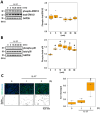
Time-dependent changes in p38 phosphorylation and ERK1/2 nuclear localization following IL-17 stimulation in normal human epidermal keratinocyte (NHEK) cells. (A,B) Immunoblot analyses of phospho-/total-ERK1/2 (A) and phospho-/total-p38 (B) after IL-17 treatment. NHEK cells were treated with 100 ng/mL IL-17 and cell lysates were collected at 0, 5, 10, 20, 30, and 60 min after treatment. Signal intensities were quantified and are presented as box plots. Phosphorylated protein levels were normalized to total protein levels. Each data point is represented as a black dot on the graph (right panels). Additionally, Glyceraldehyde 3 Phosphate Dehydrogenase (GAPDH) was detected as loading control. (C) Immunofluorescence images of ERK1/2 localization following IL-17 treatment. NHEK cells were treated with 100 ng/mL IL-17 for 0, 1, and 2 h. Cells were stained with green (ERK1/2) and blue (Hoechst) fluorescent dyes (upper panels). Green/blue co-localization signals were extracted and displayed in the lower panels. Quantified data of images were normalized to the non-treated group and presented as box plots (right panel). Each data point is represented as a black dot. Statistical significance was determined by Dunnett’s test (* p < 0.05, n = 3 to 4) compared to the zero-time control.
Figure 5
Figure 5
Effects of betulinic acid (BA) and ursolic acid (UA) on IL-17-induced p38 phosphorylation. Immunoblot images of phospho-/total-p38 in normal human epidermal keratinocyte (NHEK) cells treated with BA and UA in the presence of IL-17. NHEK cells were pretreated with 0.1 and 0.3 μM of BA or UA, followed by 100 ng/mL of IL-17 for 10 min. Signal intensities were quantified and are presented as box plots (right panel). Phosphorylated protein levels were normalized by the total protein levels. The BA and UA data are shown in red and blue, respectively. The control group (white) was treated with dimethyl sulfoxide (DMSO) only in the experiment. Each data point is represented as a black dot. Glyceraldehyde 3 Phosphate Dehydrogenase (GAPDH) was used as a loading control. Statistical significance was determined by Dunnett’s test (* p < 0.05, n = 4) compared to the IL-17-treated group (gray).
Figure 6
Figure 6
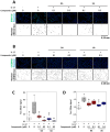
Effects of betulinic acid (BA) and ursolic acid (UA) on IL-17-induced nuclear localization of ERK1/2 and C/EBPβ. (A,B) Immunofluorescence images of ERK1/2 (A) and C/EBPβ (B) localization following treatment with IL-17 in the presence of BA and UA. Normal human epidermal keratinocyte (NHEK) cells were pretreated with 0.1 and 0.3 μM of BA or UA, followed by 100 ng/mL of IL-17 for 2 h (A) or 3 h (B). Cells were stained with green ((A) ERK1/2; (B) C/EBPβ) and blue (Hoechst) fluorescent dyes (upper panels). Green/blue co-localization signals were extracted and shown in the lower panels. Quantified data of (A) and (B) were normalized to the non-treated group and presented as box plots ((C) and (D), respectively). The control groups (white) were treated with dimethyl sulfoxide (DMSO) in all experiments. Each data point is represented as a black dot. Statistical significance was determined by Dunnett’s test (* p < 0.05, n = 4) compared to the IL-17-treated group (gray).
Figure 7
Figure 7
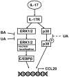
Conceptual model of the effects of betulinic acid (BA) and ursolic acid (UA) on IL-17 signaling pathways. In normal human epidermal keratinocyte (NHEK) cells, extracellular IL-17 acts on the IL-17 receptor (IL-17R). The signaling pathway downstream of IL-17R leading to CCL20 expression is broadly divided into two pathways: the p38 pathway and the ERK1/2-C/EBPβ axis. Pentacyclic triterpenoids BA and UA can significantly influence the ERK1/2-C/EBPβ axis by inhibiting ERK1/2 nuclear localization (T-shaped arrow), whereas their effect on the p38 pathway is minimal (dotted T-shaped arrow).

Similar articles

References

    1. Zhou X., Chen Y., Cui L., Shi Y., Guo C. Advances in the pathogenesis of psoriasis: From keratinocyte perspective. Cell Death Dis. 2022;13:81. doi: 10.1038/s41419-022-04523-3. - DOI - PMC - PubMed
    1. Fratton Z., Giovannini I., Zabotti A., Errichetti E. Skin and nail predictors of psoriatic arthritis development: A holistic overview integrating epidemiological and physiopathological data. J. Clin. Med. 2024;13:6880. doi: 10.3390/jcm13226880. - DOI - PMC - PubMed
    1. Armstrong A.W., Read C. Pathophysiology, clinical presentation, and treatment of psoriasis: A review. JAMA. 2020;323:1945–1960. doi: 10.1001/jama.2020.4006. - DOI - PubMed
    1. Nograles K.E., Zaba L.C., Guttman-Yassky E., Fuentes-Duculan J., Suárez-Fariñas M., Cardinale I., Khatcherian A., Gonzalez J., Pierson K.C., White T.R. Th17 cytokines interleukin (IL)-17 and IL-22 modulate distinct inflammatory and keratinocyte-response pathways. Br. J. Dermatol. 2008;159:1092–1102. doi: 10.1111/j.1365-2133.2008.08769.x. - DOI - PMC - PubMed
    1. Amatya N., Garg A.V., Gaffen S.L. IL-17 signaling: The yin and the yang. Trends Immunol. 2017;38:310–322. doi: 10.1016/j.it.2017.01.006. - DOI - PMC - PubMed

LinkOut - more resources