

Staphylococcus Strains in Atopic Dermatitis in Children: Toxins Production and Resistance Properties
- PMID: 40724622
- PMCID: PMC12299920
- DOI: 10.3390/life15071120
Staphylococcus Strains in Atopic Dermatitis in Children: Toxins Production and Resistance Properties
Abstract
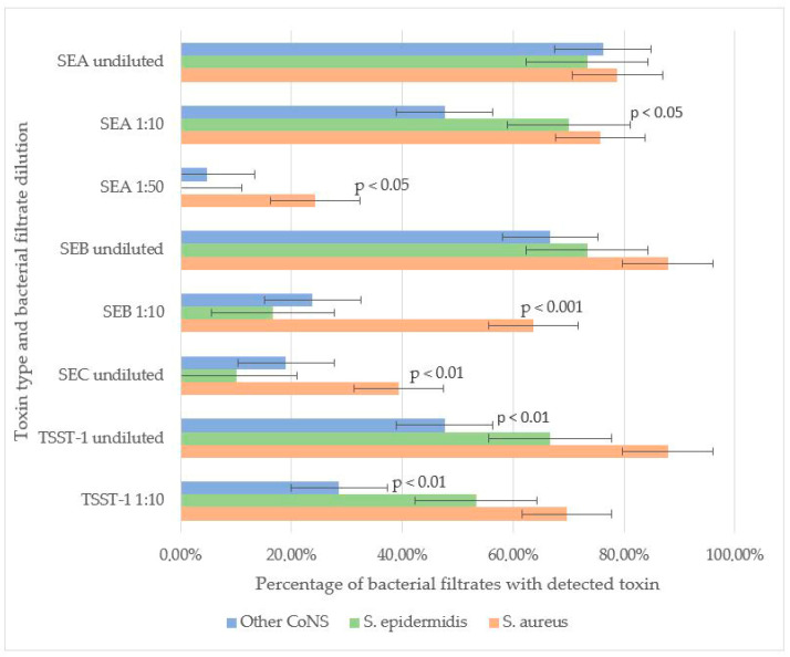
Staphylococcus spp. skin colonization is involved in the pathogenesis of atopic dermatitis (AD). While coagulase-positive Staphylococcus aureus strains are known to worsen symptoms, the role of coagulase-negative staphylococci (CoNS) remains controversial. Further research is needed to clarify the pathogenicity of CoNS in AD patients. A study involving 329 children with AD (mean age: 4.89 years) assessed the frequency of staphylococcal colonization on affected skin, along with the toxin-producing properties and antibiotic resistance of isolated strains. Mild AD: Predominantly colonized by CoNS (especially S. epidermidis). Moderate/Severe AD: Showed a significant increase in S. aureus colonization. CoNS (including S. epidermidis) could produce enterotoxins (A, B, C) and toxic shock syndrome toxin-1 (TSST-1), though less frequently than S. aureus strains. In severe AD, the number of toxin-producing CoNS strains (especially enterotoxin A producers) was higher than in mild AD, and the number of non-toxin-producing strains was lower. CoNS exhibited higher resistance rates than S. aureus. Methicillin-resistant S. epidermidis (MRSE): 23.4%. Methicillin-resistant S. aureus (MRSA): 1.27%. CoNS may contribute to AD pathogenesis through toxin production (exacerbating inflammation) and antibiotic resistance (limiting treatment options). Severe AD may involve a synergistic effect between S. aureus and toxin-producing CoNS.
Keywords: Staphylococcus aureus; Staphylococcus epidermidis; antibiotic resistance; atopic dermatitis; children; enterotoxins; toxic shock syndrome toxin-1.
Conflict of interest statement
Author Oksana Osipenko was employed by the company Family Medical Center LLC. The remaining authors declare that the research was conducted in the absence of any commercial or financial relationships that could be construed as a potential conflict of interest.
Figures


Similar articles
-
Atopic dermatitis pediatric patients show high rates of nasal and intestinal colonization by methicillin-resistant Staphylococcus aureus and coagulase-negative staphylococci.BMC Microbiol. 2024 Jan 29;24(1):42. doi: 10.1186/s12866-023-03165-5. BMC Microbiol. 2024. PMID: 38287251 Free PMC article.
-
A molecular comparative study of intestinal colonization with Staphylococcus aureus between pediatric inpatients and outpatients of different age groups.Microbiol Spectr. 2025 Jul;13(7):e0239424. doi: 10.1128/spectrum.02394-24. Epub 2025 May 16. Microbiol Spectr. 2025. PMID: 40377306 Free PMC article.
-
Epidemiology of staphylococci species and their antimicrobial-resistance among patients with wound infection in Ethiopia: a systematic review and meta-analysis.J Glob Antimicrob Resist. 2022 Jun;29:483-498. doi: 10.1016/j.jgar.2021.10.025. Epub 2021 Nov 18. J Glob Antimicrob Resist. 2022. PMID: 34801740
-
Bacteriophage infection drives loss of β-lactam resistance in methicillin-resistant Staphylococcus aureus.Elife. 2025 Jul 10;13:RP102743. doi: 10.7554/eLife.102743. Elife. 2025. PMID: 40637714 Free PMC article.
-
Prevalence and odds of Staphylococcus aureus carriage in atopic dermatitis: a systematic review and meta-analysis.Br J Dermatol. 2016 Oct;175(4):687-95. doi: 10.1111/bjd.14566. Epub 2016 Jul 5. Br J Dermatol. 2016. PMID: 26994362
References
-
- Bay L., Barnes C.J., Fritz B.G., Ravnborg N., Ruge I.F., Halling-Sønderby A.-S., Søeborg S.R., Langhoff K.H., Lex C., Hansen A.J., et al. Unique dermal bacterial signature differentiates atopic dermatitis skin from healthy. Msphere. 2025;10:e0015625. doi: 10.1128/msphere.00156-25. - DOI - PMC - PubMed
LinkOut - more resources
Full Text Sources
Miscellaneous
