. 2025 Jul 9;26(14):6581.
doi: 10.3390/ijms26146581.

Neuroinflammation Based Neurodegenerative In Vitro Model of SH-SY5Y Cells-Differential Effects on Oxidative Stress and Insulin Resistance Relevant to Alzheimer's Pathology

Affiliations

Neuroinflammation Based Neurodegenerative In Vitro Model of SH-SY5Y Cells-Differential Effects on Oxidative Stress and Insulin Resistance Relevant to Alzheimer's Pathology

Csenge Böröczky et al. Int J Mol Sci. .

Abstract

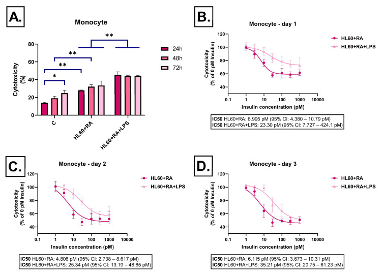
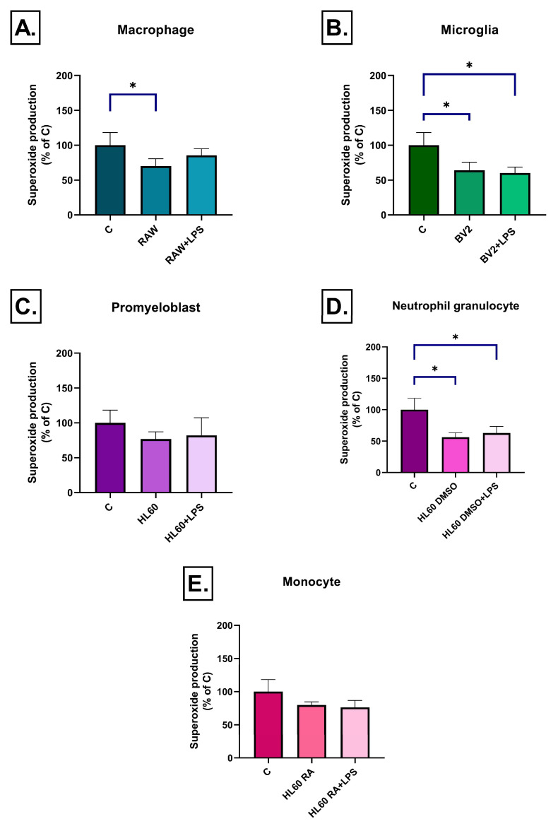
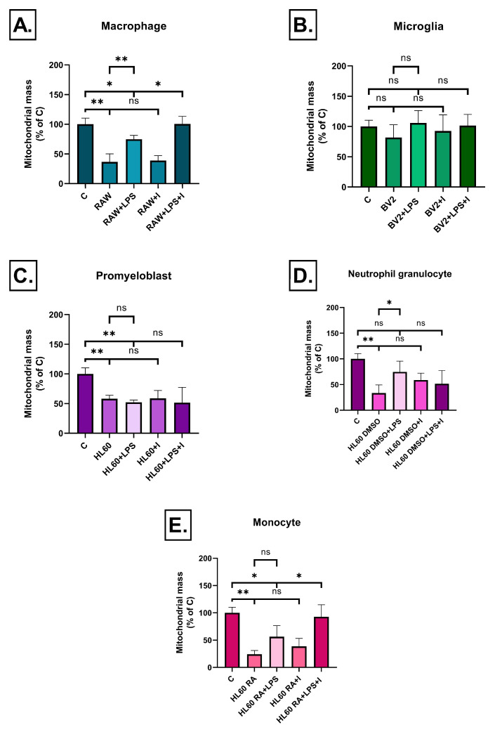
Neuroinflammation is a key process in Alzheimer's disease (AD). We aimed to examine the development and evaluation of a comprehensive in vitro model that captures the complex interplay between neurons and immune cell types. Retinoic acid-differentiated SH-SY5Y neuroblastoma cells exposed to LPS-conditioned media (CM) from RAW264.7 macrophages, BV2 microglia, and HL60 promyelocytic cells differentiated into neutrophil- or monocyte-like phenotypes were analyzed. The effects of CM containing inflammatory factors on neuronal viability and function were systematically evaluated. Neuronal oxidative stress, mitochondrial function, autophagy and protein aggregates were analyzed. The involvement of insulin resistance was studied by assaying glucose uptake and determining its IC50 values for cell viability improvement and GSK3β phosphorylation. After short-term exposure (3 h), most inflammatory CMs induced peroxide production in neurons, with the strongest effect observed in media from DMSO- or RA-differentiated HL60 cells. Mitochondrial membrane potential was markedly reduced by LPS-stimulated BV2 and HL60-derived CMs. Prolonged exposure (72 h) revealed partial normalization of oxidative stress and mitochondrial membrane potential. Glucose uptake was significantly impaired in cells treated with LPS-activated RAW264.7, BV2, and DMSO-differentiated HL60 cell media, while insulin partially rescued this effect, except for the CM of BV2 cells. Notably, insulin IC50 increased dramatically under LPS-treated BV2 cells induced inflammation (35 vs. 198 pM), confirming the development of insulin resistance. Immune cell-specific inflammation causes distinct effects on neuronal oxidative stress, mitochondrial function, protein aggregation, insulin signaling and viability. LPS-activated BV2-derived CM best recapitulates AD-related pathology, offering a relevant in vitro model for further studies.

Keywords: Alzheimer’s disease; insulin resistance; microglia; neuroinflammation.


Conflict of interest statement

The authors declare no conflicts of interest.

Figures

Figure 1
Figure 1
Effect of RAW264.7-derived conditioned media on LDH release in SH-SY5Y cells after 24 h, 48 h, and 72 h of treatment. Cells were exposed to conditioned media derived from RAW264.7 cells, either unstimulated or stimulated with LPS, and treated with insulin (0–1000 pM). LDH release was measured as a marker of cytotoxicity and expressed as a percentage of total cell lysis, normalized to spontaneous and maximal LDH release. Data are presented as % cytotoxicity. Panel (A) shows a bar chart representing baseline cytotoxicity levels at 24, 48, and 72 h without insulin treatment. Panels (BD) show the effect of insulin (0–1000 pM) on cytotoxicity. In all cases, cytotoxicity is expressed as a percentage relative to the 0 pM insulin condition. C: control; LPS: lipopolysaccharide, * p < 0.05; ** p < 0.01.
Figure 2
Figure 2
Effect of BV-2-derived conditioned media on LDH release in SH-SY5Y cells after 24 h, 48 h, and 72 h of treatment. Cells were exposed to conditioned media derived from BV-2 cells, either unstimulated or stimulated with LPS, and treated with insulin (0–1000 pM). LDH release was measured as a marker of cytotoxicity and expressed as a percentage of total cell lysis, normalized to spontaneous and maximal LDH release. Data are presented as % cytotoxicity. Panel (A) shows a bar chart representing baseline cytotoxicity levels at 24, 48, and 72 h without insulin treatment. Panels (BD) show the effect of insulin (0–1000 pM) on cytotoxicity. In all cases, cytotoxicity is expressed as a percentage relative to the 0 pM insulin condition. C: control; LPS: lipopolysaccharide, * p < 0.05; ** p < 0.01.
Figure 3
Figure 3
Effect of HL-60-derived conditioned media on LDH release in SH-SY5Y cells after 24 h, 48 h, and 72 h of treatment. Cells were exposed to conditioned media derived from HL-60 cells, either unstimulated or stimulated with LPS, and treated with insulin (0–1000 pM). LDH release was measured as a marker of cytotoxicity and expressed as a percentage of total cell lysis, normalized to spontaneous and maximal LDH release. Data are presented as % cytotoxicity. Panel (A) shows a bar chart representing baseline cytotoxicity levels at 24, 48, and 72 h without insulin treatment. Panels (BD) show the effect of insulin (0–1000 pM) on cytotoxicity. In all cases, cytotoxicity is expressed as a percentage relative to the 0 pM insulin condition. C: control; LPS: lipopolysaccharide, * p < 0.05; ** p < 0.01.
Figure 4
Figure 4
Effect of dimethyl sulfoxide-differentiated HL-60-derived conditioned media on LDH release in SH-SY5Y cells after 24 h, 48 h, and 72 h of treatment. Cells were exposed to conditioned media derived from dimethyl sulfoxide-differentiated HL-60 cells, either unstimulated or stimulated with LPS, and treated with insulin (0–1000 pM). LDH release was measured as a marker of cytotoxicity and expressed as a percentage of total cell lysis, normalized to spontaneous and maximal LDH release. Data are presented as % cytotoxicity. Panel (A) shows a bar chart representing baseline cytotoxicity levels at 24, 48, and 72 h without insulin treatment. Panels (BD) show the effect of insulin (0–1000 pM) on cytotoxicity. In all cases, cytotoxicity is expressed as a percentage relative to the 0 pM insulin condition. C: control; LPS: lipopolysaccharide, DMSO: dimethyl-sulfoxide, * p < 0.05; ** p < 0.01.
Figure 5
Figure 5
Effect of all-trans retinoic acid-differentiated HL-60-derived conditioned media on LDH release in SH-SY5Y cells after 24 h, 48 h, and 72 h of treatment. Cells were exposed to conditioned media derived from all-trans retinoic acid-differentiated HL-60 cells, either unstimulated or stimulated with LPS, and treated with insulin (0–1000 pM). LDH release was measured as a marker of cytotoxicity and expressed as a percentage of total cell lysis, normalized to spontaneous and maximal LDH release. Data are presented as % cytotoxicity. Panel (A) shows a bar chart representing baseline cytotoxicity levels at 24, 48, and 72 h without insulin treatment. Panels (BD) show the effect of insulin (0–1000 pM) on cytotoxicity. In all cases, cytotoxicity is expressed as a percentage relative to the 0 pM insulin condition. C: control; LPS: lipopolysaccharide, RA: all-trans retinoic acid, * p < 0.05; ** p < 0.01.
Figure 6
Figure 6
Peroxide production in SH-SY5Y cells after 3 h of treatment with conditioned media from various immune cell types (AE), measured by 2′,7′-dichlorofluorescin diacetate (DCFDA) fluorescence. LPS-stimulated conditioned media induced a significant increase in ROS levels compared to the control in all cell line groups. Data are expressed as a percentage of the control. C: control, LPS: lipopolysaccharide, RA: all-trans retinoic acid, DMSO: dimethyl sulfoxide, * p < 0.05; ** p < 0.01.
Figure 7
Figure 7
Peroxide production in SH-SY5Y cells after 72 h of treatment with conditioned media from various immune cell types (AE), measured by 2′,7′-dichlorofluorescin diacetate (DCFDA) fluorescence. Compared to the 3 h time point, peroxide levels were normalized by 72 h in most groups, shown by no significant differences relative to the control, with the exception of the RA-differentiated HL60 cell medium. Data are expressed as a percentage of the control. C: control, LPS: lipopolysaccharide, RA: all-trans retinoic acid, DMSO: dimethyl sulfoxide, * p < 0.05.
Figure 8
Figure 8
Superoxide production in SH-SY5Y cells after 3 h of treatment with conditioned media from various immune cell types (AE), measured by dihydroethidium (HE) fluorescence. A significant increase in superoxide production was observed only in response to the LPS-stimulated conditioned media of RAW264.7 and DMSO-differentiated HL60 cells, while no significant changes were detected in the other groups. Data are expressed as a percentage of the control. C: control, LPS: lipopolysaccharide, RA: all-trans retinoic acid, DMSO: dimethyl sulfoxide, * p < 0.05.
Figure 9
Figure 9
Superoxide production in SH-SY5Y cells after 72 h of treatment with conditioned media from various immune cell types (AE), measured by dihydroethidium (HE) fluorescence. A significant decrease in superoxide production was observed in response to LPS-stimulated conditioned media from RAW264.7, BV2, and DMSO-differentiated HL-60 cells, while no significant changes were detected in the other two groups. Data are expressed as a percentage of the control. C: control, LPS: lipopolysaccharide, RA: all-trans retinoic acid, DMSO: dimethyl sulfoxide, * p < 0.05.
Figure 10
Figure 10
Mitochondrial membrane potential in SH-SY5Y cells after 3 h of treatment with conditioned media from various immune cell types (AE), measured by JC-1 fluorescence. A significant increase in the green/red fluorescence ratio was observed in all LPS-treated groups, indicating mitochondrial depolarization compared to the control. Data are expressed as a percentage of the control. C: control, LPS: lipopolysaccharide, RA: all-trans retinoic acid, DMSO: dimethyl sulfoxide, * p < 0.05; ** p < 0.01.
Figure 11
Figure 11
Mitochondrial membrane potential in SH-SY5Y cells after 72 h of treatment with conditioned media from various immune cell types (AE), measured by JC-1 fluorescence. Compared to the 3-h time point, the green/red fluorescence ratio returned to baseline levels in all groups by 72 h, with no significant differences relative to the control. Data are expressed as a percentage of the control. C: control, LPS: lipopolysaccharide, RA: all-trans retinoic acid, DMSO: dimethyl sulfoxide.
Figure 12
Figure 12
Mitochondrial mass in SH-SY5Y cells after 72 h of treatment with conditioned media from various immune cell types (AE), measured by MitoTracker fluorescence. Changes in mitochondrial signal intensity varied between treatment groups. Data are expressed as a percentage of the control. C: control, LPS: lipopolysaccharide, RA: all-trans retinoic acid, DMSO: dimethyl sulfoxide, * p < 0.05; ** p < 0.01, ns: not significant.
Figure 13
Figure 13
Glucose uptake into SH-SY5Y cells after 72 h of treatment with conditioned media from various immune cell types (AE), measured by 2-NBDG fluorescence. Reduced glucose uptake was seen in the case of LPS-treated RAW264.7, BV2 and DMSO differentiated HL60 cells. Insulin treatment reversed it only in the case of RAW264.7 cells. Data are expressed as a percentage of the control. C: control, LPS: lipopolysaccharide, RA: all-trans retinoic acid, DMSO: dimethyl sulfoxide, ** p < 0.01, ns: not significant.
Figure 14
Figure 14
Autophagic vacuole accumulation in SH-SY5Y cells after 72 h of treatment with conditioned media from various immune cell types (AE), measured by acridine orange staining. Fluorescence data were normalized to cell number, and control values were subtracted from each treatment group. Representative spectra are shown alongside bar graphs of the corresponding area under the curve (ΔAUC) values. C: control, LPS: lipopolysaccharide, RA: all-trans retinoic acid, DMSO: dimethyl sulfoxide, ** p < 0.01.
Figure 14
Figure 14
Autophagic vacuole accumulation in SH-SY5Y cells after 72 h of treatment with conditioned media from various immune cell types (AE), measured by acridine orange staining. Fluorescence data were normalized to cell number, and control values were subtracted from each treatment group. Representative spectra are shown alongside bar graphs of the corresponding area under the curve (ΔAUC) values. C: control, LPS: lipopolysaccharide, RA: all-trans retinoic acid, DMSO: dimethyl sulfoxide, ** p < 0.01.
Figure 15
Figure 15
Phosphorylation of GSK3β in SH-SY5Y cells after 72 h of treatment with conditioned media derived from BV2 microglia, with or without liraglutide, followed by insulin stimulation (1 h, 1–1000 pM). C: control, LPS: lipopolysaccharide.
Figure 16
Figure 16
Thioflavin S staining of protein aggregates in SH-SY5Y cells after treatment with conditioned media from BV2 microglia, with or without LPS and/or insulin (1000 pM) treatment. (A) Representative fluorescence images showing thioflavin S (green), DAPI (blue), and merged channels. (B) Quantification of protein aggregation expressed as the ratio of thioflavin S to DAPI fluorescence. Conditioned media from LPS-stimulated BV2 cells induced a significant increase in amyloid aggregation that was reversed by insulin treatment. C: control, LPS: lipopolysaccharide, I: insulin, * p < 0.05; ns: not significant.

References

    1. Kwon H.S., Koh S.H. Neuroinflammation in neurodegenerative disorders: The roles of microglia and astrocytes. Transl. Neurodegener. 2020;9:42. doi: 10.1186/s40035-020-00221-2. - DOI - PMC - PubMed
    1. Kinney J.W., Bemiller S.M., Murtishaw A.S., Leisgang A.M., Salazar A.M., Lamb B.T. Inflammation as a central mechanism in Alzheimer’s disease. Alzheimers Dement. 2018;4:575–590. doi: 10.1016/j.trci.2018.06.014. - DOI - PMC - PubMed
    1. Parbo P., Ismail R., Sommerauer M., Stokholm M.G., Hansen A.K., Hansen K.V., Amidi A., Schaldemose J.L., Gottrup H., Braendgaard H., et al. Does inflammation precede tau aggregation in early Alzheimer’s disease? A PET study. Neurobiol. Dis. 2018;117:211–216. doi: 10.1016/j.nbd.2018.06.004. - DOI - PubMed
    1. Muller L., Di Benedetto S. Neuroimmune crosstalk in chronic neuroinflammation: Microglial interactions and immune modulation. Front. Cell. Neurosci. 2025;19:1575022. doi: 10.3389/fncel.2025.1575022. - DOI - PMC - PubMed
    1. Brown G.C., Neher J.J. Microglial phagocytosis of live neurons. Nat. Rev. Neurosci. 2014;15:209–216. doi: 10.1038/nrn3710. - DOI - PubMed

MeSH terms

LinkOut - more resources