. 2025 Jul 10;26(14):6603.
doi: 10.3390/ijms26146603.

Radiation-Sensitive Nano-, Micro-, and Macro-Gels and Polymer Capsules for Use in Radiotherapy Dosimetry

Affiliations

Radiation-Sensitive Nano-, Micro-, and Macro-Gels and Polymer Capsules for Use in Radiotherapy Dosimetry

Michał Piotrowski et al. Int J Mol Sci. .

Abstract

This work introduces an original approach to the manufacturing of ionizing radiation-sensitive systems for radiotherapy applications-dosimetry. They are based on the Fricke dosimetric solution and the formation of macro-gels and capsules, and nano- and micro-gels. The reaction of ionic polymers, such as sodium alginate, with Fe and Ca metal ions is employed. Critical polymer concentration (c*) is taken as the criterion. Reaction of ionic polymers with metal ions leads to products related to c*. Well below c*, nano- and micro-gels may form. Above c*, macro-gels and capsules can be prepared. Nano- and micro-gels containing Fe in the composition can be used for infusion of a physical gel matrix to prepare 2D or 3D dosimeters. In turn, macro-gels can be formed with Fe ions crosslinking polymer chains to obtain radiation-sensitive hydrogels, so-called from wall-to-wall, serving as 3D dosimeters. The encapsulation process can lead to capsules with Fe ions serving as 1D dosimeters. This work presents the concept of manufacturing various gel structures, their main features and manufacturing challenges. It proposes new directions of research towards novel dosimeters.

Keywords: 3D dosimeter; Fricke gel dosimeter; ionizing radiation; macro-gels; micro-gels; radiotherapy dosimeter.


Conflict of interest statement

The authors declare no conflicts of interest.

Figures

Figure 1
Figure 1
The concept of the Fricke 3D gel dosimeter. 3D gel matrix containing (i) nano- or micro-gels made of alginate (chitosan or other ionic polymer) crosslinked with Fe ions (also with, e.g., Ca ions), (ii) a macro-gel made of alginate (chitosan or other ionic polymer) crosslinked with Fe ions (also with, e.g., Ca ions), and (iii) a single capsule made of alginate (chitosan or other ionic polymer) crosslinked with Fe ions (also with, e.g., Ca ions). Below is a scheme illustrating the course of the study related to the manufacturing of the Fricke 3D radiotherapy gel dosimeter materials as capsules, macro-gels, and containing nano- or micro-gels in structure.
Figure 2
Figure 2
Alginate capsules obtained by dropping the sodium alginate polymer solution of a low viscosity (Sigma) into the Fricke solution containing CaCl2 of 0% (A,F), 1% (B,G), 1.5% (C,H), 2.5% (D,I), and 3.5% (E,J). The concentration of the sodium alginate solution was as follows: 1.7% (AE), 3.5% (FJ).
Figure 3
Figure 3
Alginate capsules obtained by dropping the sodium alginate polymer solution of medium viscosity (Sigma) into the Fricke solution containing CaCl2 of 0% (A,D), 1% (B,E), and 1.5% (C,F). The concentration of the sodium alginate solution was as follows: 1.7% (AC), 3.5% (DF).
Figure 4
Figure 4
Alginate capsules obtained by dropping the sodium alginate polymer solution (medium viscosity, Heppe) into the Fricke solution containing CaCl2 of 0% (A,F), 1% (B,G), 1.5% (C,H), 2.5% (D,I), and 3.5% (E,J). The concentration of the sodium alginate solution was as follows: 1.7% (AE), 3.5% (FJ).
Figure 5
Figure 5
Stability of the capsules made in a reaction of 3.5% alginate (low viscosity Sigma) with Fricke solution (FAS: 1 mM, XO: 0.165 mM, H2SO4: 50 mM) containing 3.5% CaCl2 by dropwise addition of sodium alginate solution into the Fricke solution with Ca+2 (A). The capsules stored in the Fricke solution either in a refrigerator at approximately 4 °C or in a cabinet at approximately 21–23 °C (without access to daylight). Top of the figure contains photographs of the capsules during storage (A). The graph (B) corresponds to the color values of the capsules read with ImageJ software (v. 1.53j, National Institutes of Health, Bethesda, MD, USA) over time of storage (mean values of circular ROIs covering the entire capsules).
Figure 6
Figure 6
(A): Stability of alginate capsules containing Fricke solution. A mean value of color for the photographs taken with a microscope (Delta Optical Smart 5MP PRO digital microscope, Delta Optical, Nowe Osiny, Poland) versus storage time at approximately 25 °C with access to laboratory light, removed from Fricke solution, for the capsules formed with Heppe medium-viscosity and Sigma low-viscosity alginates. Beneath the graph are the photographs (B) of the capsules taken at 0–150 min after formation (first row: capsules made of Heppe and second row: capsules made of Sigma alginate). The capsules were made in a reaction of 3.5% alginate with Fricke solution (FAS: 1 mM, XO: 0.165 mM, H2SO4: 50 mM) containing 3.5% CaCl2 by dropwise addition of sodium alginate solution into the Fricke solution with Ca+2.
Figure 7
Figure 7
Response of alginate capsules containing Fricke solution to irradiation with X-rays of a TrueBeam medical accelerator (Varian, Palo Alto, CA, USA): mean value of color for the photographs taken with a microscope (Delta Optical Smart 5MP PRO digital microscope) versus absorbed dose (A) and example photographs of non-irradiated and irradiated capsules (B). The capsules were made of Heppe and a low-viscosity Sigma alginate in a reaction of 3.5% alginate with Fricke solution (FAS: 1 mM, XO: 0.165 mM, H2SO4: 50 mM) containing 3.5% CaCl2 by dropwise addition of sodium alginate solution into the Fricke solution with Ca+2. The capsules were immersed (0 Gy) and removed from the Fricke solution (20 Gy) and not immersed in the solution after irradiation. The photographs of capsules are magnified: ×38. The values in A are averages for 3–6 capsules and were calculated from approximately 50,000 pixels.
Figure 8
Figure 8
Content of Fe (A) and Ca (B) per 1 kg of capsules (average weight of one capsule: 0.034 g) in relation to reaction time between sodium alginate solution (Sigma and Heppe) and Fricke solution (FAS: 1 mM, H2SO4: 50 mM, XO: 0.165 mM) containing 3.5% CaCl2. Measurements were performed with the aid of ICP-OES. The reaction was carried out at a temperature of approximately 23 °C. Throughout the experiment, the contents of the beaker were stirred with a magnetic stirrer at a speed of approximately 450 rpm.
Figure 9
Figure 9
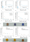
Examination of diffusion of Fe (A,C,F) and Ca (B,D,G) from alginate capsules (average weight of one capsule: 0.034 g) made by reaction of sodium alginate solution (Sigma) (3.5%) with Fricke solution (FAS: 1 mM, H2SO4: 50 mM, XO: 0.165 mM; reaction time = 40 min) containing 3.5 CaCl2 (A,B), different concentrations of FAS (C,D), and different compounds containing Fe (F,G) (concentration of each compound: 1 mM). In (E,H), photographs of capsules made of different concentrations of FAS and different compounds containing Fe, respectively, are shown. The capsules were immersed in 3.5 mL of re-distilled water. Measurements were performed with the aid of ICP-OES. For (A,B), the water with the capsules was changed after 48 h of diffusion, for (C,D), the water was changed after 180 min, and for (F,G), after 120 min.
Figure 10
Figure 10
Response to irradiation of alginate capsules with Fricke solution in Pluronic F-127 or gelatine matrix. (A): Irradiation set up. Samples in cuvettes of 4.7 cm height and 1 cm optical path are immersed in water (water phantom, W-1-DD1-2, GeVero Co.); the water phantom is on the bench of the TrueBeam accelerator (Varian). (B): Top view of the samples in the water phantom. (C,D): Samples before and after irradiation (20 Gy) for the dosimeters with Pluronic F-127 (C) and gelatine (D) matrix; in (D), a cuvette with gelatine matrix and capsule containing Fricke dosimeter without XO dye is also shown (G.-XO) for comparison. P. and G. denote Pluronic F-127 and gelatine, respectively; P.W. and G.W. denote Pluronic F-127 and gelatine matrices, respectively, with capsules that were washed (for 2 days) before immersion in the matrices and after irradiation (20 Gy). (E,F): Profiles along the samples as indicated by green dashed line in (C). The values are for the green channel of RGB color space.
Figure 10
Figure 10
Response to irradiation of alginate capsules with Fricke solution in Pluronic F-127 or gelatine matrix. (A): Irradiation set up. Samples in cuvettes of 4.7 cm height and 1 cm optical path are immersed in water (water phantom, W-1-DD1-2, GeVero Co.); the water phantom is on the bench of the TrueBeam accelerator (Varian). (B): Top view of the samples in the water phantom. (C,D): Samples before and after irradiation (20 Gy) for the dosimeters with Pluronic F-127 (C) and gelatine (D) matrix; in (D), a cuvette with gelatine matrix and capsule containing Fricke dosimeter without XO dye is also shown (G.-XO) for comparison. P. and G. denote Pluronic F-127 and gelatine, respectively; P.W. and G.W. denote Pluronic F-127 and gelatine matrices, respectively, with capsules that were washed (for 2 days) before immersion in the matrices and after irradiation (20 Gy). (E,F): Profiles along the samples as indicated by green dashed line in (C). The values are for the green channel of RGB color space.
Figure 11
Figure 11
Performance of alginate capsules containing Fricke dosimeter embedded in Pluronic F-127 and gelatine matrices. (A): Pluronic F-127 matrix, c CaCl2 = 1.5%; (B): gelatine matrix, c CaCl2 = 1.5%; (C): gelatine matrix, c CaCl2 = 0%; (D,E): change of size of alginate capsule in Pluronic F-127 (D) and gelatine (E) matrix over time of storage at room temperature (~23 °C). Capsules formed in a reaction of 3.5% sodium alginate with Fricke solution: 1 mM FAS, 50 mM H2SO4, and 0.165 mM XO containing 0–3.5% CaCl2. Black bars in (AC) denote the distance of 0.75 mm. Dashed lines indicate the positions where the diameter was measured.
Figure 11
Figure 11
Performance of alginate capsules containing Fricke dosimeter embedded in Pluronic F-127 and gelatine matrices. (A): Pluronic F-127 matrix, c CaCl2 = 1.5%; (B): gelatine matrix, c CaCl2 = 1.5%; (C): gelatine matrix, c CaCl2 = 0%; (D,E): change of size of alginate capsule in Pluronic F-127 (D) and gelatine (E) matrix over time of storage at room temperature (~23 °C). Capsules formed in a reaction of 3.5% sodium alginate with Fricke solution: 1 mM FAS, 50 mM H2SO4, and 0.165 mM XO containing 0–3.5% CaCl2. Black bars in (AC) denote the distance of 0.75 mm. Dashed lines indicate the positions where the diameter was measured.
Figure 12
Figure 12
Performance of alginate capsules containing Fricke dosimeter embedded in Pluronic F-127 and gelatine matrices. (A,B) correspond to the capsules treated with poly-L-lysine and immersed in the Pluronic F-127 and gelatine matrices, respectively. (C,D) are for the capsules immersed in Pluronic F-127 (C) and gelatine matrices (D) at different ionic strength—concentrations of NaCl. Capsules formed in a reaction of 3.5% sodium alginate with Fricke solution: 1 mM FAS, 50 mM H2SO4, and 0.165 mM XO containing 3.5% CaCl2.
Figure 12
Figure 12
Performance of alginate capsules containing Fricke dosimeter embedded in Pluronic F-127 and gelatine matrices. (A,B) correspond to the capsules treated with poly-L-lysine and immersed in the Pluronic F-127 and gelatine matrices, respectively. (C,D) are for the capsules immersed in Pluronic F-127 (C) and gelatine matrices (D) at different ionic strength—concentrations of NaCl. Capsules formed in a reaction of 3.5% sodium alginate with Fricke solution: 1 mM FAS, 50 mM H2SO4, and 0.165 mM XO containing 3.5% CaCl2.
Figure 13
Figure 13
Irradiation of alginate capsules. Phantom SP34 (RW3 plates) (A) and a poly(methyl methacrylate) template with holes and capsules inside the holes (B).

Similar articles

References

    1. Slotman B.J., Solberg T.D., Verellen D., editors. Extracranial Stereotactic Radiotherapy and Radiosurgery. Taylor & Francis; Abingdon-on-Thames, UK: 2005.
    1. Trifiletti D.M., Chao S.T., Sahgal A., Sheehan J.P., editors. Stereotactic Radiosurgery and Stereotactic Body Radiation Therapy. A Comprehensive Guide. Springer Nature; Basel, Switzerland: 2019.
    1. Swiss Society of Radiobiology and Medical Physics . Quality Control of Medical Electron Accelerators. Recommendations Number 11. Swiss Society of Radiobiology and Medical Physics; Geneva, Switzerland: 2015.
    1. Fricke H., Morse S. The chemical action of Roentgen rays on dilute ferrosulphate solutions as a measure of dose. Am. J. Roent. Radium Ther. Nucl. Med. 1927;18:430–432.
    1. Gore J.C., Kang Y.S., Schultz R.J. Measurement of radiation dose distributions by nuclear magnetic resonance (NMR) imaging. Phys. Med. Biol. 1984;29:1189–1199. doi: 10.1088/0031-9155/29/10/002. - DOI - PubMed

LinkOut - more resources