Review
. 2025 Jul 10;26(14):6613.
doi: 10.3390/ijms26146613.

The Role of Lactylation in Virus-Host Interactions

Affiliations
Review

The Role of Lactylation in Virus-Host Interactions

Gejie Zhao et al. Int J Mol Sci. .

Abstract

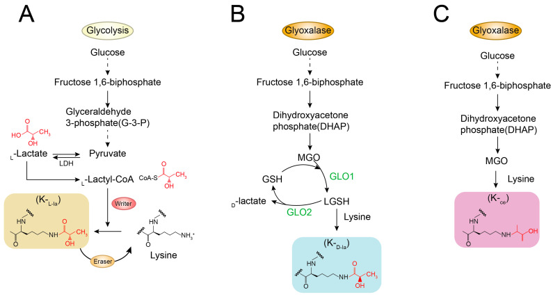
Lactylation, a novel form of post-translational modifications (PTMs) of protein, particularly within histone proteins, has recently gained attention for its role in regulating gene expression and cellular processes. In recent years, lactylation has been widely studied in cancer, immune diseases, neurological diseases, cardiovascular diseases, metabolic diseases, etc. Increasing evidence now suggests that lactylation also plays a significant role in the host's innate immune response to viruses. Lactylation influences fundamental cellular functions, including transcriptional regulation, signal transduction, cell proliferation and differentiation. It affects protein behavior by modulating their function, stability, subcellular localization and interactions. Studies have shown that many viral infections promote lactate production through enhanced glycolysis, a process that facilitates viral replication. Given that innate immunity serves as the host's first line of defense against pathogenic invasion, understanding how lactylation regulates antiviral responses offers promising avenues for the development of diagnostic tools and therapeutic strategies against viral diseases. In this review, we provide a comprehensive overview of recent research on the role of lactylation in viral-host interactions.

Keywords: innate immunity; lactylation; post-translational modifications (PTMs); viral infection; virus.


Conflict of interest statement

The authors declare no conflicts of interest.

Figures

Figure 1
Figure 1
Overview of lactate production and its role in lactylation. Under aerobic conditions: Pyruvate is oxidized to acetyl-CoA by the pyruvate dehydrogenase complex (PDH). Acetyl-CoA then enters the tricarboxylic acid (TCA) cycle, followed by oxidative phosphorylation, ultimately generating ATP. Under anaerobic conditions: Pyruvate is reduced to lactate by the lactate dehydrogenase (LDH). Additionally, cells can import extracellular lactate via monocarboxylate transporters (MCTs). Intracellular lactate can be converted to lactyl-CoA, which serves as a substrate for lactylation modifications, thereby facilitating the lactylation of both histone and non-histone proteins. Inspired by the lactate metabolism framework described by Li et al. [14].
Figure 2
Figure 2
Three isomers of lysine lactylation. (A). KL-la: Forms enzymatically when glycolytically derived L-lactate (produced by LDH from pyruvate) is converted to lactyl-CoA and conjugated to lysine residues. (B). Lysine D-lactylation (KD-la): The stereoisomer of KL-la. Forms via spontaneous reaction between lysine residues and D-lactate generated by the glyoxalase pathway (processing the glycolytic byproduct methylglyoxal, MGO). (C). N-ε-(carboxyethyl)lysine (Kce): An adduct formed by the direct, non-enzymatic reaction of the highly reactive glycolytic byproduct methylglyoxal (MGO) with lysine residues. MGO is a highly reactive byproduct of glycolysis that can react with a variety of protein residues, including cysteine, arginine, and lysine. N-ε-(carboxyethyl)lysine (Kce), formed by the reaction with lysine, has been detected in cells, although its levels are lower than those of MGO-derived arginine modifications. Inspired by the lactylation isomer framework described by Zhang, D. et al. [19]. The dotted line indicates that the process involves multiple intermediate steps, which have been omitted here for simplicity. The solid line represents a direct reaction. Different colors are used solely for visual distinction.
Figure 3
Figure 3
Lactylation regulates virus-associated signaling pathways. (1) cGAS Inhibition: Viral infection promotes the lactate-dependent lactylation of cGAS via the cGAS: AARS1/2 complex. This modification suppresses cGAS DNA-sensing activity, reducing cGAMP synthesis and inhibiting innate immunity. (2) PRRSV Immune Evasion: PRRSV infection elevates cellular lactate, driving the lactylation of Heat Shock Protein Family A Member 6 (HSPA6). Lactylated HSPA6 disrupts TRAF3-IKKε complex formation, blocking IFN-β production and compromising antiviral responses. (3) RLR Pathway Suppression: During infection by 5′ppp-dsRNA, SeV, or VSV, glycolytic lactate accumulation enables direct lactate binding to MAVS. This interaction prevents MAVS mitochondrial oligomerization, inhibiting RLR signaling activation. (4) CSFV Antiviral Defense: CSFV-induced lactate activates histone H2B lactylation (H2BK16la). This modification recruits karyopherin subunit alpha 2 (KPNA2) to facilitate p65/NF-κB nuclear translocation, stimulating IFN-λ expression and suppressing viral replication. (5) SVA Immune Evasion: Senecavirus A (SVA) enhances its replication by exploiting glycolysis-induced lactate production to disrupt the MAVS-RIG-I interaction and suppress RIG-I-like receptor (RLR) signaling. Inspired by the virus-associated signaling pathway framework described by Zhang, W., Li, H., Zhu, W., Pang, Y. et al. [11,21,30,31,32].

Similar articles

References

    1. Ren H., Tang Y., Zhang D. The emerging role of protein L-lactylation in metabolic regulation and cell signalling. Nat. Metab. 2025;7:647–664. doi: 10.1038/s42255-025-01259-0. - DOI - PubMed
    1. Cui H., Xie N., Banerjee S., Ge J., Jiang D., Dey T., Matthews Q.L., Liu R.M., Liu G. Lung Myofibroblasts Promote Macrophage Profibrotic Activity through Lactate-induced Histone Lactylation. Am. J. Respir. Cell Mol. Biol. 2021;64:115–125. doi: 10.1165/rcmb.2020-0360OC. - DOI - PMC - PubMed
    1. Wang J., Peng M., Oyang L., Shen M., Li S., Jiang X., Ren Z., Peng Q., Xu X., Tan S., et al. Mechanism and application of lactylation in cancers. Cell Biosci. 2025;15:76. doi: 10.1186/s13578-025-01415-9. - DOI - PMC - PubMed
    1. Krishnan S., Nordqvist H., Ambikan A.T., Gupta S., Sperk M., Svensson-Akusjärvi S., Mikaeloff F., Benfeitas R., Saccon E., Ponnan S.M., et al. Metabolic Perturbation Associated with COVID-19 Disease Severity and SARS-CoV-2 Replication. Mol. Cell Proteom. 2021;20:100159. doi: 10.1016/j.mcpro.2021.100159. - DOI - PMC - PubMed
    1. Meng X., Zhu Y., Yang W., Zhang J., Jin W., Tian R., Yang Z., Wang R. HIF-1α promotes virus replication and cytokine storm in H1N1 virus-induced severe pneumonia through cellular metabolic reprogramming. Virol. Sin. 2024;39:81–96. doi: 10.1016/j.virs.2023.11.010. - DOI - PMC - PubMed

LinkOut - more resources