. 2025 Jul 11;26(14):6651.
doi: 10.3390/ijms26146651.

Identification of Genes Linked to Meniscal Degeneration in Osteoarthritis: An In Silico Analysis

Affiliations

Identification of Genes Linked to Meniscal Degeneration in Osteoarthritis: An In Silico Analysis

Aliki-Alexandra Papageorgiou et al. Int J Mol Sci. .

Abstract

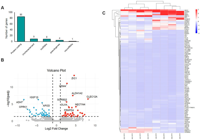
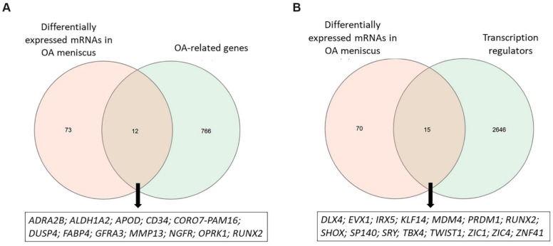
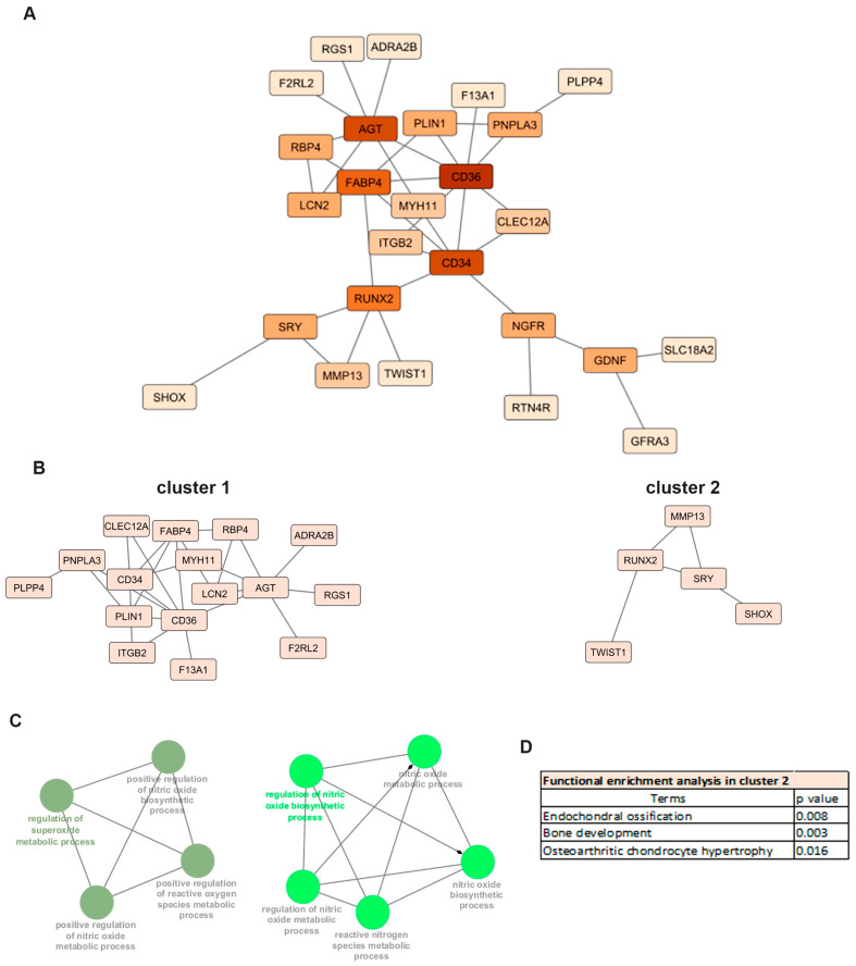
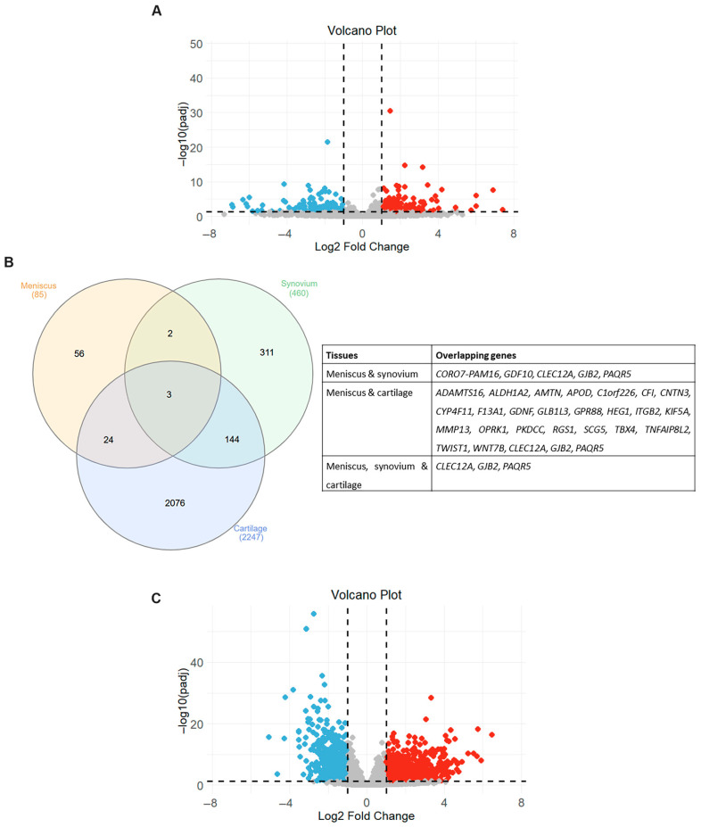
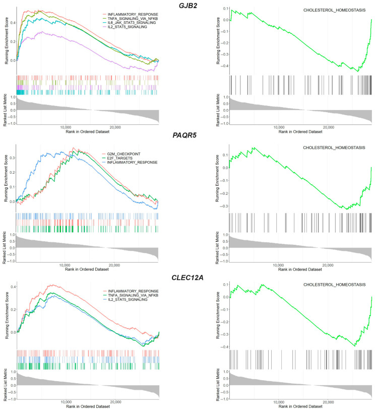
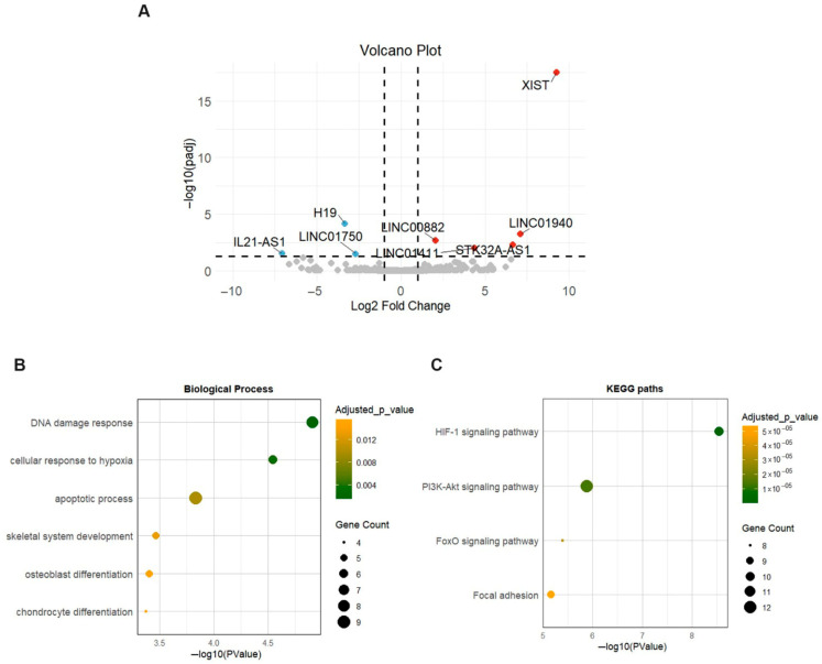
Meniscal degradation is considered a driver of osteoarthritis (OA) progression, but the underlying mechanisms leading to age-related meniscus degeneration remain unknown. This study aimed to identify key genes and pathways involved in meniscal degradation through a computational analysis. Gene expression profiles were obtained from the Gene Expression Omnibus (GEO) database. Differential expression gene (DEG) analysis was performed using DESeq2 accompanied by functional enrichment analysis, protein-protein interaction (PPI) and clustering analysis. Additionally, gene set enrichment analysis (GSEA) was performed. A total of 85 mRNAs (DEMs) and 8 long non-coding RNAs (DE LncRNAs) were found to be differentially expressed in OA meniscus tissues. Among 85 DEMs, 12 genes were found to be known OA-related genes, whereas 15 genes acted as transcription regulators, including RUNX2 and TBX4, which were identified as effector genes for OA. Enrichment analysis revealed the implication of DEMs in cartilage-degradation-related processes, including inflammatory pathways, lipid metabolism, extracellular matrix organization and superoxide/nitric oxide metabolic processes. Target genes of DE lncRNAs were found to be involved in chondrocyte differentiation and pathways related to cartilage degradation. A comparative analysis of meniscus, synovium and cartilage datasets identified three genes (GJB2, PAQR5 and CLEC12A) as being differentially expressed across all three OA-affected tissues, which were implicated in inflammatory and cholesterol metabolism processes. Our results support that shared mechanisms lead to meniscal and cartilage degradation during OA progression, providing further insights into the processes underlying OA pathogenesis and potential therapeutic targets for knee OA.

Keywords: in silico analysis; meniscus; osteoarthritis; transcriptome.


Conflict of interest statement

The authors declare no conflicts of interest.

Figures

Figure 1
Figure 1
mRNA expression profile of OA meniscus: (A) Different biotypes of genes differentially expressed between OA and healthy meniscus (adjusted p-value < 0.05). (B) Volcano plot showing the differentially expressed mRNAs between OA and healthy meniscus (adjusted p-value < 0.05; absolute log2 fold change > 1). Red dots indicate significantly upregulated mRNAs and blue dots significantly downregulated mRNAs. Gray dots represent mRNAs without significant differential expression. (C) Heatmap indicating the expression patterns of OA and healthy meniscus.
Figure 2
Figure 2
Identification of OA-related DEMs and transcription regulators. (A) Venn diagram showing the overlap between differentially expressed mRNAs observed in OA meniscus and OA-related genes obtained by Open Targets platform. (B) Venn diagram showing the overlap between differentially expressed mRNAs observed in OA meniscus and transcription regulators obtained by the AnimalTFDB database.
Figure 3
Figure 3
Protein–protein interaction (PPI) analysis: (A) PPI network of differentially expressed mRNAs in OA meniscus. The nodes represent the target genes and the edges indicate both functional and physical protein associations. A high confidence score of 0.400 was defined as the minimum interaction score to construct the PPI network. (B) Sub-networks after MCL clustering. (C) Enrichment analyses of cluster 1 using the ClueGo plugin of Cytoscape. (D) Functional enrichment analysis of cluster 2 using g:Profiler.
Figure 4
Figure 4
Overlapping differentially expressed genes (DEGs) in different joint components. (A) Volcano plot showing differentially expressed mRNAs between OA and healthy synovium samples. Red dots indicate significantly upregulated mRNAs and blue dots significantly downregulated mRNAs (adjusted p-value < 0.05 and |log2 fold change| > 1). Gray dots represent mRNAs without significant differential expression. (B) Venn diagram and table showing the overlapping DEGs in three OA-affected joint tissues. (C) Volcano plot showing differentially expressed mRNAs between OA and healthy cartilage samples. Red dots indicate significantly upregulated mRNAs and blue dots significantly downregulated mRNAs (adjusted p-value < 0.05 and |log2 fold change| > 1). Gray dots represent mRNAs without significant differential expression.
Figure 5
Figure 5
GSEA enrichment analysis of overlapping genes GJB2, PAQR5 and CLEC12A. GSEA enrichment plot showing the biological processes in which the overlapping genes were involved.
Figure 6
Figure 6
Expression profile of lncRNAs in OA meniscus. (A) Volcano plot showing differentially expressed lncRNAs between OA and healthy meniscus samples. Red dots indicate significantly upregulated lncRNAs and blue dots significantly downregulated lncRNAs (adjusted p-value < 0.05 and |log2 fold change| > 1). Gray dots represent lncRNAs without significant differential expression. (B,C) Dot plots show the enriched biological processes and KEGG pathways of target genes of differentially expressed lncRNAs. Dot size indicates the number of targets enriched in each pathway and the dot color reflects the adjusted p-value.

Similar articles

References

    1. Li H.Z., Liang X.Z., Sun Y.Q., Jia H.F., Li J.C., Li G. Global, regional, and national burdens of osteoarthritis from 1990 to 2021: Findings from the 2021 global burden of disease study. Front. Med. 2024;11:1476853. doi: 10.3389/fmed.2024.1476853. - DOI - PMC - PubMed
    1. Kong H., Wang X.Q., Zhang X.A. Exercise for Osteoarthritis: A Literature Review of Pathology and Mechanism. Front. Aging Neurosci. 2022;14:854026. doi: 10.3389/fnagi.2022.854026. - DOI - PMC - PubMed
    1. Yao Q., Wu X., Tao C., Gong W., Chen M., Qu M., Zhong Y., He T., Chen S., Xiao G. Osteoarthritis: Pathogenic signaling pathways and therapeutic targets. Signal Transduct. Target. Ther. 2023;8:56. doi: 10.1038/s41392-023-01330-w. - DOI - PMC - PubMed
    1. Kloppenburg M., Namane M., Cicuttini F. Osteoarthritis. Lancet. 2025;405:71–85. doi: 10.1016/S0140-6736(24)02322-5. - DOI - PubMed
    1. Sanchez-Lopez E., Coras R., Torres A., Lane N.E., Guma M. Synovial Inflammation in Osteoarthritis Progression. Nat. Rev. Rheumatol. 2022;18:258–275. doi: 10.1038/s41584-022-00749-9. - DOI - PMC - PubMed

LinkOut - more resources