. 2025 Jul 11;26(14):6653.
doi: 10.3390/ijms26146653.

Simulated Microgravity Attenuates Stretch Sensitivity of Mechanically Gated Channels in Rat Ventricular Myocytes

Affiliations

Simulated Microgravity Attenuates Stretch Sensitivity of Mechanically Gated Channels in Rat Ventricular Myocytes

Andrey S Bilichenko et al. Int J Mol Sci. .

Abstract

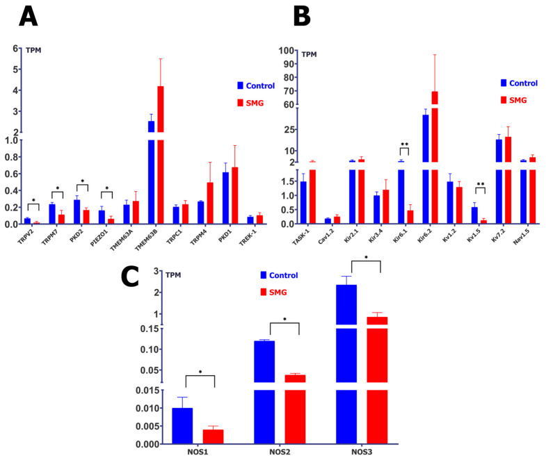
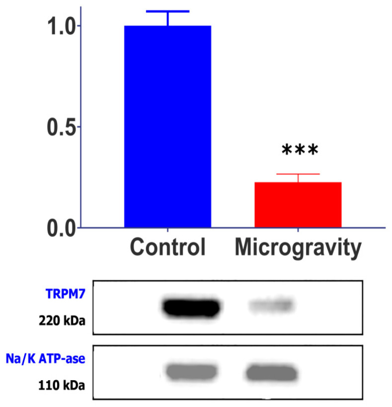
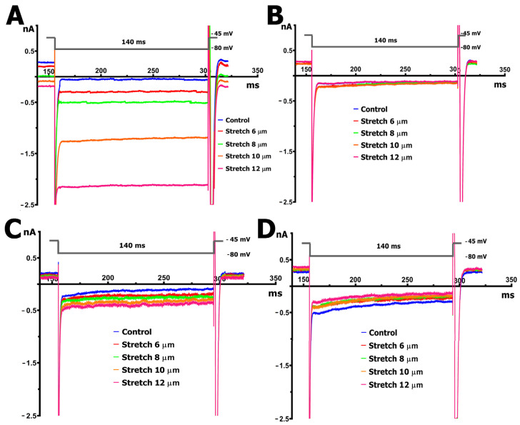
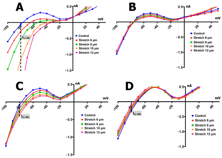
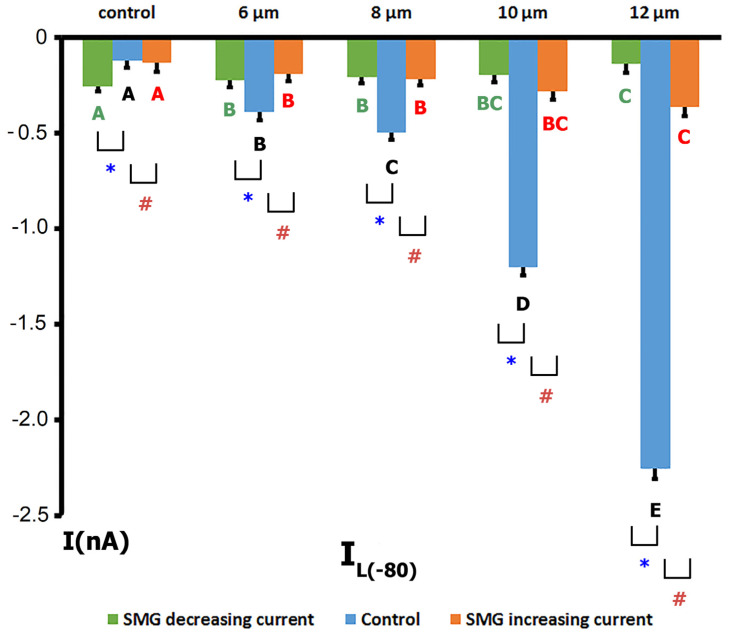
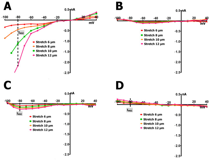
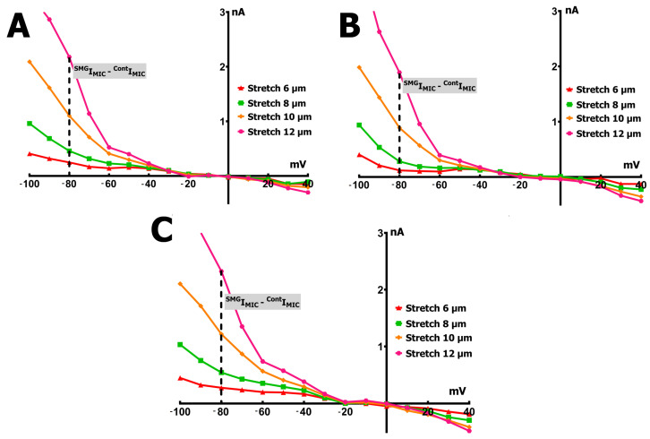
Cardiomyocytes, similarly to cells in various tissues, are responsive to mechanical stress of all types, which is reflected in the significant alterations to their electrophysiological characteristics. This phenomenon, known as mechanoelectric feedback, is based on the work of mechanically gated channels (MGCs) and mechano-sensitive channels (MSCs). Since microgravity (MG) in space, as well as simulated microgravity (SMG), changes the morphological and physiological properties of the heart, it was assumed that this result would be associated with a change in the expression of genes encoding MGCs and MSCs, leading to a change in the synthesis of channel proteins and, ultimately, a change in channel currents during cell stretching. In isolated ventricular cardiomyocytes of rats exposed to SMG for 14 days, the amount of MGCs and MSCs gene transcripts was studied using the RNA sequencing method by normalizing the amount of "raw" reads using the Transcripts Per Kilobase Million (TPM) method. Changes in the level of channel protein, using the example of the MGCs TRPM7, were assessed by the Western blot method, and changes in membrane ion currents in the control and during cardiomyocyte stretching were assessed by the patch-clamp method in the whole-cell configuration. The data obtained demonstrate that SMG results in a multidirectional change in the expression of genes encoding various MGCs and MSCs. At the same time, a decrease in the TPM of the MGCs TRPM7 gene leads to a decrease in the amount of TRPM7 protein. The resulting redistribution in the synthesis of most channel proteins leads to a marked decrease in the sensitivity of the current through MGCs to cell stretching and, ultimately, to a change in the functioning of the heart.

Keywords: TRPM7; mechanosensitive channels; rat; simulated microgravity; stretch; ventricular myocytes.


Conflict of interest statement

The authors declare no conflicts of interest.

Figures

Figure 1
Figure 1
The effect of simulated microgravity (SMG) on transcript levels of mechanically gated channels (MGCs), mechanosensitive channels (MSCs), and nitric oxide synthases (NOS). Gene expression was quantified and normalized using the TPM (Transcripts Per Kilobase Million) method. (A): Expression of MGC-related transcripts under SMG. Blue bars represent control (n = 7; red bars represent SMG-treated samples (n = 4). * p < 0.05; all other comparisons not significant (NS). (B) Expression of MSC-related transcripts under SMG. Blue bars—control (n = 7); red bars—SMG (n = 4). ** p < 0.001; all other comparisons NS (Kv and Kir channels in this figure are presented under the category of MSCs due to their reported mechanosensitive modulation or physiological relevance to membrane responses under stretch, though they are not classical MGCs). (C) Expression of NOS isoforms under SMG. Blue bars—control (n = 7); red bars—SMG (n = 4). * p < 0.05.
Figure 2
Figure 2
Cardiomyocyte TRPM7 channel protein levels normalized to housekeeping protein (α-subunit of Na+/K+-ATPase, validated as stable under SMG conditions by RNA-seq analysis, p = 0.89) in control animals (n = 11) and animals subjected to SMG (n = 8). *** p < 0.001 compared to control.
Figure 3
Figure 3
Original recordings of the membrane currents at a holding potential of −45 mV with a 140 ms step to −80 mV before cell stretching (blue curve) and upon stretching by 6 (red curve), 8 (green curve), 10 (orange curve), and 12 (pink curve) µm. (A) Shift in the currents towards negative values upon the graded stretching of cardiomyocytes from control rats. (B) No change in the currents upon the graded stretching of cardiomyocytes from rats exposed to SMG. (C) Decreased shift in the currents towards negative values (compared to control rats in “A”) upon the graded stretching of cardiomyocytes from rats exposed to SMG. (D) Slight shift in the currents towards positive values upon the graded stretching of cardiomyocytes from rats exposed to SMG.
Figure 4
Figure 4
Voltage dependence of the IL recorded in ventricular cardiomyocytes from control rats and rats exposed to SMG, before and during graded mechanical stretching. IL reflects the net IMIC, comprising contributions from IMGC and IMSC. Blue curves indicate control (unstretched) conditions; red, green, orange, and pink curves represent stretches of 6, 8, 10, and 12 µm, respectively. (A) Control group: IL increases progressively with stretch, especially at negative potentials, indicating the robust activation of mechanosensitive currents. (B) SMG group—type I response: no significant change in IL across stretch levels, indicating loss of stretch sensitivity. (C) SMG group—type II response: modest increase in IL with stretch, but significantly attenuated compared to control. (D) SMG group—type III response: IL decreases with stretch, suggesting a paradoxical or reversed response to mechanical loading.
Figure 5
Figure 5
Bar graph showing mean values of IL at −80 mV in ventricular cardiomyocytes from control rats (blue bars; n = 9) and rats exposed to SMG. IL values were measured under baseline conditions and during graded mechanical stretching (6, 8, 10, and 12 µm). The SMG group is subdivided into two response phenotypes: cells with a slight increase in IL (orange bars; n = 8, corresponding to Figure 3C) and those with a decrease in IL (green bars; n = 6, corresponding to Figure 3D). Letters (A–E) above bars of the same color indicate statistically significant differences between stretch levels within a group (p < 0.01); identical letters denote no significant difference (NS). Asterisks (*) and hash marks (#) indicate statistically significant differences between control and SMG groups at the corresponding stretch level (p < 0.01).
Figure 6
Figure 6
Voltage dependence of the net mechanically induced current (IMIC) in ventricular cardiomyocytes from control rats and those exposed to SMG, calculated as the difference between late currents recorded under stretch (SIL) and control conditions (CIL): IMIC = SILCIL. Each panel shows IMIC at −80 mV across increasing stretch amplitudes: 6 µm (red), 8 µm (green), 10 µm (orange), and 12 µm (pink). (A) Control group: robust, progressive increase in inward IMIC with stretch, indicating normal mechanosensitivity. (B) SMG group—type I response: complete absence of stretch-induced current, reflecting loss of mechanotransductive response. (C) SMG group—type II response: attenuated IMIC compared to controls, suggesting reduced mechanosensitivity. (D) SMG group—type III response: paradoxical positive IMIC values, indicating reversed response to mechanical stretching.
Figure 7
Figure 7
Difference in net IMIC between cardiomyocytes from SMG rats and control rats, plotted as SIMICCIMIC across membrane potentials. Curves represent stretch amplitudes of 6 µm (red), 8 µm (green), 10 µm (orange), and 12 µm (pink). (A) Type I response: no significant change in IMIC across stretch levels, indicating preserved absence of mechanosensitive current in SMG cardiomyocytes. (B) Type II response: consistently reduced IMIC in SMG cells compared to controls, reflecting diminished stretch sensitivity. (C) Type III response: positive shift in IMIC difference, consistent with reversed (paradoxical) response to stretch under SMG conditions.

Similar articles

References

    1. Ravens U. Mechano-electric feedback and arrhythmias. Prog. Biophys. Mol. Biol. 2003;82:255–266. doi: 10.1016/S0079-6107(03)00026-9. - DOI - PubMed
    1. Craelius W., Chen V., el-Sherif N. Stretch activated ion channels in ventricular myocytes. Biosci. Rep. 1988;8:407–414. doi: 10.1007/BF01121637. - DOI - PubMed
    1. Kamkin A., Kiseleva I., Wagner K.D., Leiterer K.P., Theres H., Scholz H., Günther J., Lab M.J. Mechano-electric feedback in right atrium after left ventricular infarction in rats. J. Mol. Cell. Cardiol. 2000;32:465–477. doi: 10.1006/jmcc.1999.1091. - DOI - PubMed
    1. Kiseleva I., Kamkin A., Wagner K.D., Theres H., Ladhoff A., Scholz H., Günther J., Lab M.J. Mechanoelectric feedback after left ventricular infarction in rats. Cardiovasc. Res. 2000;45:370–378. doi: 10.1016/S0008-6363(99)00361-2. - DOI - PubMed
    1. Kamkin A., Kiseleva I., Isenberg G. Stretch-activated currents in ventricular myocytes: Amplitude and arrhythmogenic effects increase with hypertrophy. Cardiovasc. Res. 2000;48:409–420. doi: 10.1016/S0008-6363(00)00208-X. - DOI - PubMed

LinkOut - more resources