Review
. 2025 Jul 11;26(14):6667.
doi: 10.3390/ijms26146667.

Retinoic Acid Induced 1 and Smith-Magenis Syndrome: From Genetics to Biology and Possible Therapeutic Strategies

Affiliations
Review

Retinoic Acid Induced 1 and Smith-Magenis Syndrome: From Genetics to Biology and Possible Therapeutic Strategies

Jasmine Covarelli et al. Int J Mol Sci. .

Abstract

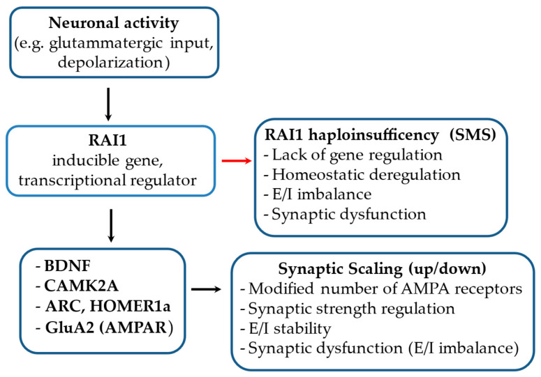
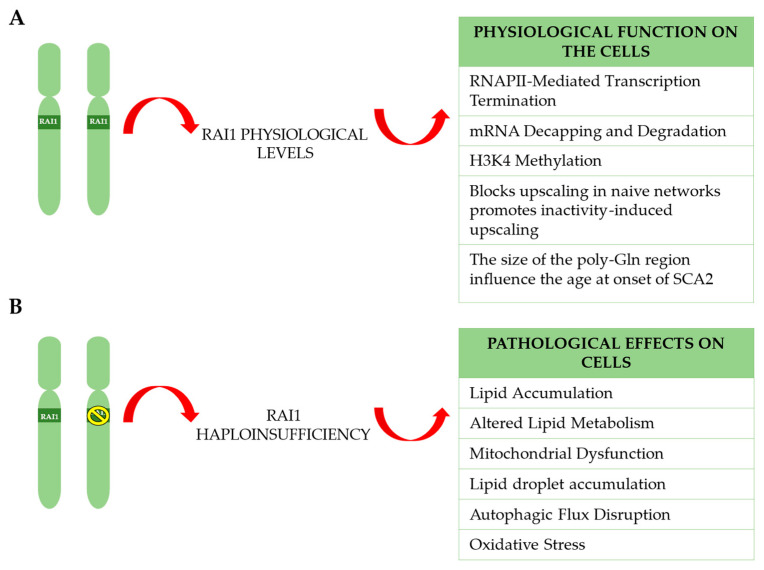
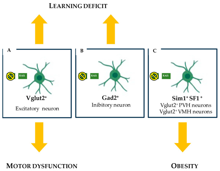
Haploinsufficiency disorders are genetic diseases caused by reduced gene expression, leading to developmental, metabolic, and tumorigenic abnormalities. The dosage-sensitive Retinoic Acid Induced 1 (RAI1) gene, located within the 17p11.2 region, is central to the core features of Smith--Magenis syndrome (SMS) and Potocki--Lupski syndrome (PTLS), caused by the reciprocal microdeletions and microduplications of this region, respectively. SMS and PTLS present contrasting phenotypes. SMS is characterized by severe neurobehavioral manifestations, sleep disturbances, and metabolic abnormalities, and PTLS shows milder features. Here, we detail the molecular functions of RAI1 in its wild-type and haploinsufficiency conditions (RAI1+/-), as studied in animal and cellular models. RAI1 acts as a transcription factor critical for neurodevelopment and synaptic plasticity, a chromatin remodeler within the Histone 3 Lysine 4 (H3K4) writer complex, and a regulator of faulty 5'-capped pre-mRNA degradation. Alterations of RAI1 functions lead to synaptic scaling and transcriptional dysregulation in neural networks. This review highlights key molecular mechanisms of RAI1, elucidating its role in the interplay between genetics and phenotypic features and summarizes innovative therapeutic approaches for SMS. These data provide a foundation for potential therapeutic strategies targeting RAI1, its mRNA products, or downstream pathways.

Keywords: HD; PTLS; RAI1; SMS.


Conflict of interest statement

The authors declare no conflicts of interest.

Figures

Figure 1
Figure 1
Genomic rearrangements by non-allelic homologous recombination (NAHR) between directly oriented LCRs. The figure illustrates the three different mechanisms by which structural genomic rearrangements can occur, leading to imbalances such as microdeletion, microduplication, and acentric chromosomes. (A) Interchromosomal NAHR. This schematic shows the potential NAHR between two homologous chromosome pairs. The presence of LCRs (striped and hatched) with high homology can lead to one allele slipping onto the other, resulting in a 1:1 unbalanced segregation of alleles. In this case, two chromosomes remain normal, while the other two comprise one that is microdeleted and the other microduplicated. (B) Intrachromosomal NAHR. The image shows recombination between two chromatids. For proper alignment of the LCR there is a possibility that the chromosome, instead of aligning correctly, may fold back on itself due to the high homology downstream of the LCR. This leads to the formation of two unbalanced chromosomes. (C) Intrachromatid NAHR. This image shows the final possibility, where NAHR occurs within the same chromatid. Due to the high homology of the LCR, the chromosome folds onto itself to facilitate the binding between the LCR located further downstream. This complex and rare structural rearrangement results in one normal chromosome, one carrying the microdeletion, and one acentric chromosome. N: normal; del: deletion; dup: duplication; ace: acentric.
Figure 2
Figure 2
The genomic architecture of the RAI1 gene (A) and chromosome region 17p11.2 (B) with the mechanisms underlying deletions leading to genomic imbalances. (A) A schematic representation of the human RAI1 (OMIM #607642, NM_030665.4 GRCh38/hg38), which spans ~130 kb on chromosome 17, from positions 17,681,458 to 17,811,453 (UCSC Genome Browser). The coding exons (3, 4, 5, and part of 6) are shown in dark green, while the non-coding exons (1–2 and part of 6) are in light green. The 5′ and 3′ untranslated regions (UTRs) are represented by thick gray lines. Translation starts in exon 3, which encodes the majority (~97%) of the RAI1 protein. Exon 3 is also a mutational hotspot, often affected in RAI1-related disorders. (B) The genomic organization of the 17p11.2 region, illustrating the location of RAI1 (green box) and the flanking low-copy repeats (LCRs; green and white stripes) and SMS–Repetitive Element (SMS-REPs: SMS-REPD, SMS-REPM, and SMS-REPP; shown as a brick-pattern motif; D = distal, M = middle, P = proximal). These highly homologous repeat elements mediate NAHR events, leading to recurrent or uncommon deletions. The most frequent rearrangement—the common deletion—occurs between SMS-REPD and SMS-REPP. Other deletion types (uncommon or large) involve recombination between different combinations of LCRs (LCR17pA–D). The orientation of the region is indicated from telomere to centromere.
Figure 3
Figure 3
Diagrammatic representation of chromosome 17, highlighting the common deletion at the 17p11.2 locus. From left to right, the following elements are depicted: the G-band ideogram of human chromosome 17; and the schematic illustration of the Smith–Magenis syndrome region with OMIM related genes—emphasizing the nature of SMS as a contiguous gene syndrome and the contribution of multiple gene deletion to the complex and systemic phenotypic manifestations of the syndrome. The red box indicates the location of the RAI1 gene in relation to other genes within the 17p11.2 region.
Figure 4
Figure 4
RAI1 Protein Structure. This figure illustrates the structural organization of the RAI1 protein, highlighting its several key functional domains: N-terminal polyglutamine-rich tract (Poly-Q), polyserine-rich domain (Poly-S), bipartite nuclear localization signal (NLS), sometimes considered as two distinct domains, second polyserine-rich tract (Poly-S), nucleosome-binding domain (NBD), and C-terminal plant homeodomain (PHD).
Figure 5
Figure 5
Diagram of the potential involvement of RAI1 in synaptic scaling. Neuronal activity induces the expression of RAI1, an activity-dependent transcriptional regulator that modulates genes involved in synaptic plasticity and the excitatory–inhibitory (E/I) balance, including BDNF, CAMK2A, ARC, HOMER1a, and GluA2. These targets contribute to synaptic scaling, a homeostatic mechanism that adjusts synaptic strength in response to prolonged changes in network activity. RAI1 haploinsufficiency may disrupt this regulatory pathway, leading to synaptic dysfunctions associated with neurodevelopmental disorders. The red arrow highlights the altered impact of RAI1 haploinsufficiency on synaptic scaling.
Figure 6
Figure 6
Schematic overview of RAI1 activity in physiological conditions and following haploinsufficiency. (A) Under physiological conditions, both alleles of the RAI1 gene are expressed. RAI1 regulates multiple key cellular processes, including RNA polymerase II (RNAPII)-mediated transcription termination, mRNA decapping and degradation, and H3K4 methylation, all of which are essential for proper gene expression control. In neural networks, RAI1 blocks activity-independent upscaling and promotes inactivity-induced synaptic scaling. Additionally, a polymorphism in RAI1′s polyglutamine (poly-GLn) tract may modulate the age at onset in SCA2. (B) RAI1 haploinsufficiency disrupts these cellular functions, leading to a cascade of pathological cellular effects, particularly in neurons, including lipid accumulation, altered lipid metabolism, mitochondrial dysfunction, lipid droplet accumulation, autophagic flux disruption, and oxidative stress. The red arrows indicate the flow from genetic dosage to cellular outcomes in both physiological and pathological contexts.
Figure 7
Figure 7
Neuronal roles of RAI1 in motor dysfunction and obesity in Smith–Magenis syndrome. This schematic illustrates RAI1 expression across key neuronal subtypes implicated in SMS phenotypes: excitatory Vglut2+ neurons, associated with motor delay, cognitive deficits, and obesity (AC); inhibitory Gad2+ neurons (B), linked to cognitive deficits; and Sim1+ and SF1+ neurons located in the paraventricular (PVH) and ventromedial (VMH) hypothalamic nuclei, which are involved in appetite regulation (C).
Figure 8
Figure 8
RAI1 deficiency impairs BDNF–TRKB signaling. Neuronal subtypes expressing or lacking RAI1 are shown. Under normal conditions, BDNF–TRKB signaling promotes satiety. In SMS, RAI1 deficiency leads to reduced BDNF levels, decreased TRKB activation, impaired satiety signaling, hyperphagia, and obesity. RAI1 binds to promoter IV of the BDNF gene.

Similar articles

References

    1. Wilkie A.O.M. The Molecular Basis of Genetic Dominance. J. Med. Genet. 1994;31:89–98. doi: 10.1136/jmg.31.2.89. - DOI - PMC - PubMed
    1. Javed S., Selliah T., Lee Y.J., Huang W.H. Dosage-sensitive genes in autism spectrum disorders: From neurobiology to therapy. Neurosci. Biobehav. Rev. 2020;118:538–567. doi: 10.1016/j.neubiorev.2020.08.009. - DOI - PubMed
    1. Falco M., Amabile S., Acquaviva F. Rai1 Gene Mutations: Mechanisms of Smith–Magenis Syndrome. Appl. Clin. Genet. 2017;10:85–94. doi: 10.2147/TACG.S128455. - DOI - PMC - PubMed
    1. Yanagisawa T., Murayama Y., Ehara H., Goto M., Aoki M., Sekine S.I. Structural Basis of Eukaryotic Transcription Termination by the Rat1 Exonuclease Complex. Nat. Commun. 2024;15:7854. doi: 10.1038/s41467-024-52157-0. - DOI - PMC - PubMed
    1. Klama S., Hirsch A.G., Schneider U.M., Zander G., Seel A., Krebber H. A Guard Protein Mediated Quality Control Mechanism Monitors 5’-Capping of Pre-MRNAs. Nucleic Acids Res. 2022;50:11301–11314. doi: 10.1093/nar/gkac952. - DOI - PMC - PubMed

MeSH terms

LinkOut - more resources