. 2025 Jul 12;26(14):6707.
doi: 10.3390/ijms26146707.

Citrate Transporter Expression and Localization: The Slc13a5Flag Mouse Model

Affiliations

Citrate Transporter Expression and Localization: The Slc13a5Flag Mouse Model

Jan C-C Hu et al. Int J Mol Sci. .

Abstract

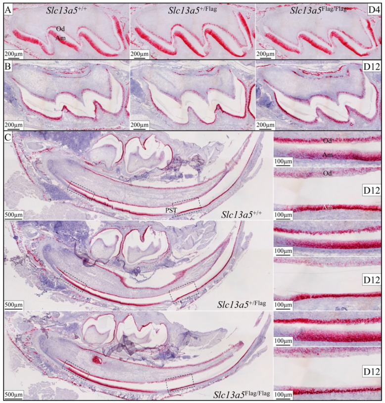
The sodium-citrate cotransporter (NaCT) plays a crucial role in citrate transport during amelogenesis. Mutations in the SLC13A5 gene, which encodes the NaCT, cause early infantile epileptic encephalopathy 25 and amelogenesis imperfecta. We analyzed developing pig molars and determined that the citrate concentrations in secretory- and maturation-stage enamel are both 5.3 µmol/g, with about 95% of the citrate being bound to mineral. To better understand how citrate might enter developing enamel, we developed Slc13a5Flag reporter mice that express NaCT with a C-terminal Flag-tag (DYKDDDDK) that can be specifically and accurately recognized by commercially available anti-Flag antibodies. The 24-base Flag coding sequence was located immediately upstream of the natural translation termination codon (TAG) and was validated by Sanger sequencing. The general development, physical activities, and reproductive outcomes of this mouse strain were comparable to those of the C57BL/6 mice. No differences were detected between the Slc13a5Flag and wild-type mice. Tooth development was extensively characterized using dissection microscopy, bSEM, light microscopy, in situ hybridization, and immunohistochemistry. Tooth formation was not altered in any detectable way by the introduction of the Flag. The Slc13a5Flag citrate transporter was observed on all outer membranes of secretory ameloblasts (distal, lateral, and proximal), with the strongest signal on the Tomes process, and was detectable in all but the distal membrane of maturation-stage ameloblasts. The papillary layer also showed positive immunostaining for Flag. The outer membrane of odontoblasts stained stronger than ameloblasts, except for the odontoblastic processes, which did not immunostain. As NaCT is thought to only facilitate citrate entry into the cell, we performed in situ hybridization that showed Ank is not expressed by secretory- or maturation-stage ameloblasts, ruling out that ANK can transport citrate into enamel. In conclusion, we developed Slc13a5Flag reporter mice that provide specific and sensitive localization of a fully functional NaCT-Flag protein. The localization of the Slc13a5Flag citrate transporter throughout the ameloblast membrane suggests that either citrate enters enamel by a paracellular route or NaCT can transport citrate bidirectionally (into or out of ameloblasts) depending upon local conditions.

Keywords: amelogenesis; enamel mineralization; protein localization.


Conflict of interest statement

The authors declare no conflicts of interest.

Figures

Figure 1
Figure 1
Generation and validation of the Slc13a5Flag knock-in mouse model. (A) The gene targeting construct used a Neo (neomycin) cassette selection marker flanked by FRT sites inserted into intron 11 of mouse Slc13a5. Note: Flp recombinase-mediated recombination occurs between these FRT sites. A Flag-tag (DYKDDDDK) coding sequence (GACTACAAAGACGATGACGACAAG) was introduced immediately upstream of the natural TAG translation termination codon in exon 12 using homologous recombination. (B) To identify Slc13a5Flag mice, the 5′ ends of the Slc13a5 and Slc13a5Flag genes were amplified from tail biopsies with a primer pair that, following PacI restriction, yielded an uncut 719 bp product from the wild type and an 840 bp knock-in product cut into 500 and 340 bp fragments that were visualized by agarose gel electrophoresis. The gel image demonstrated two wild-type mice (lanes 1 and 5), three heterozygous mice Slc13a5+/Flag (lanes 2, 4, and 6), and one homozygous Slc13a5Flag/Flag in lane 3. (C) Slc13a5+/+ and Slc13a5Flag/Flag sequence chromatograms showing 48 nucleotides replaced the TAG stop codon and added code for the 8-amino-acid Flag-tag (shown in fluorescent green) and a downstream PacI restriction site (for ease of genotyping).
Figure 2
Figure 2
Dissection microscope images of Slc13a5+/+, Slc13a5+/Flag, and Slc13a5Flag/Flag mouse incisors, molars, and hemimandibles at 7 weeks. (A) Labial view of incisors from two mice of each genotype. (B) Going clockwise from upper right are mesial, distal, lingual, and labial views of a mandibular incisor. (C) Occlusal (top), buccal, and lingual (bottom) views of molars. (D) Lingual (top) and lateral (bottom) views of a hemimandible from each genotype. No differences were observed at 7 weeks among the WT (Slc13a5+/+), heterozygous (Slc13a5+/Flag), and homozygous (Slc13a5Flag/Flag) mandibular incisors, molars, or mandible.
Figure 3
Figure 3
Backscattered scanning electron microscopy (bSEM) of Slc13a5+/+, Slc13a5+/Flag, and Slc13a5Flag/Flag 7-week mandibular incisors cross-sectioned at 1 mm increments. (A) The incisor sections are arranged in a time-forward developmental sequence from left (cervical/earliest development) to right (incisal/more advanced development). The secretory stage of enamel formation (when enamel steadily expands in cross-sectional area) is complete or nearly complete in the second column of cross-sections. By the third column, enamel formation is entirely in the maturation stage where the cross-sectional area of enamel is constant but becomes progressively more mineralized (whiter). Dentin (surrounding the radiolucent dental pulp in the center) continues to increase in cross-sectional area by the further deposition of dentin by odontoblasts lining the pulp. Dentin is less mineralized than enamel and contains characteristic dentinal tubules occupied by odontoblast processes. (B) Higher magnification images showing the dentinal tubules nearest the dentinoenamel junction (DEJ). (C) Higher magnification images of the highly mineralized enamel. The heterozygous (Slc13a5+/Flag) and homozygous (Slc13a5Flag/Flag) mandibular incisors were indistinguishable from the wild type (Slc13a5+/+).
Figure 4
Figure 4
Slc13a5+/+, Slc13a5+/Flag, and Slc13a5Flag/Flag mandibular mouse molars evaluated by bSEM. (A) Day 14 mandibular first molars immediately prior to their eruption through soft tissue into the oral cavity. The crowns are fully formed but have not yet been altered by occlusal forces. (B) Molar crowns at 7 weeks that had been functioning for ~5 weeks. The heterozygous (Slc13a5+/Flag) and homozygous (Slc13a5Flag/Flag) mouse teeth were indistinguishable from the wild type (Slc13a5+/+).
Figure 5
Figure 5
In situ hybridization shows Slc13a5 mRNA expression is not altered by the insertion of the Flag sequence. (A) Slc13a5+/+, Slc13a5+/Flag, and Slc13a5Flag/Flag Day 4 maxillary first molars, in which all ameloblasts are still in the secretory stage and show comparable patterns of Slc13a5 expression in both ameloblasts (Ams) and odontoblasts (Ods). (B) Slc13a5+/+, Slc13a5+/Flag, and Slc13a5Flag/Flag Day 12 maxillary first molars, in which all ameloblasts have transitioned into the maturation stage and show the same patterns of Slc13a5 expression in ameloblasts and odontoblasts. (C) Ameloblasts in post-secretory transition (PST) were negative for Slc13a5 mRNA expression. Mandibular cross-sections are on the left. Dashed rectangles on the left in these sections correspond to odontoblasts above early-secretory-stage ameloblasts in the higher magnification sections (top right). Dashed rectangles on the right correspond to odontoblasts above early-maturation-stage ameloblasts in the higher magnification sections (bottom right). Slc13a5+/+, Slc13a5+/Flag, and Slc13a5Flag/Flag Day 12 mandibular incisors all showed the same patterns of Slc13a5 expression.
Figure 6
Figure 6
Incisor and molar development was not altered histologically by the insertion of the Flag sequence into Slc13a5. (A) H&E-stained postnatal Day 4 (D4) maxillary first molars showing overall crown morphology (top) and higher magnification sections from dashed regions (bottom). Note: the morphologies of secretory-stage ameloblasts, odontoblasts, and the dentin and enamel layers are all comparable. (B) Comparable images of D12 maxillary first molars during the maturation stage. Overall molar shape root development, dentin, and maturation-stage enamel layers are comparable. Higher magnification panels (bottom) of the boxed areas show that the odontoblasts and maturation-stage ameloblast morphologies are all normal. (C) Longitudinal sections of D12 mandibular incisors were similar among the three genotypes (left). Higher magnifications of the odontoblasts (Od), dentin (d), enamel (e), and ameloblasts (Am) showed normal cell morphology and deposition of mineral.
Figure 7
Figure 7
Immunohistochemistry of Day 12 longitudinal sections of Slc13a5Flag/Flag (left) and wild-type (right) mandibular incisors triple-stained using PA1-984B antibodies against the Flag-tag on the NaCT (red), β-actin antibodies (green) to stain the ameloblast Tomes processes, and DAPI (blue) to stain nuclei. (A) Low-magnification views of mandibular incisors. Top: incisor sections stained for Flag (red), β-actin (green), and nuclei (blue). Bottom: incisor sections showing only Flag stain. Note: ameloblasts were positively stained for NaCT-Flag (left), with a minor background signal observed in the wild type (right, where no Flag is expressed). (B) High-magnification images of early-secretory-stage ameloblasts positive for Flag (left) and triple-stained (right). (C) Mid-secretory-stage ameloblasts stained heavily for the NaCT (left), and this signal overlapped with β-actin in the Tomes process (right). (D) The highest magnification views of the positive signal for NaCT-Flag in the Tomes processes. Note the minimal background of the positive Flag signal in the wild-type sections, which did not express the Flag protein.
Figure 8
Figure 8
Immunohistochemistry of Day 12 longitudinal sections of Slc13a5Flag/Flag (left) and wild-type (right) mandibular incisors triple-stained using 740001 antibodies against the Flag-tag on the NaCT (red), β-actin antibodies (green) to stain the ameloblast Tomes processes, and DAPI (blue) to stain nuclei. (A) Low-magnification views of entire incisors. Top: incisor sections stained for Flag (red), β-actin (green), and nuclei (blue). Bottom: incisor sections showing only Flag signal. Note: ameloblasts were positive for NaCT-Flag (left), with some background signal in the wild type (right) associated with intercellular junctions. (B) High-magnification images of early-secretory-stage ameloblasts immunostained for Flag (left) and triple-stained (right). (C) Mid-secretory-stage ameloblasts stained heavily for the NaCT (left), and this signal overlapped with β-actin in the Tomes process (right). (D) The highest magnification views of the positive signal for NaCT-Flag in the Tomes processes. Note the background of the positive Flag signal in wild-type sections (right) was higher than what was observed using the PA1-984B antibody in Figure 7 and Figure 9.
Figure 9
Figure 9
Immunohistochemistry of Day 12 longitudinal sections of Slc13a5Flag/Flag (left) and wild-type (right) mandibular incisors triple-stained using PA1-984B antibodies to stain NaCT-Flag (red), β-actin antibodies (green), and DAPI (blue) to stain nuclei. (A) Early-maturation-stage ameloblasts (lacking Tomes processes) immunostained for Flag only (left) and triple-stained (right). NaCT-Flag localizes throughout the cell membrane of the maturation-stage ameloblasts. Note the minimal background of the positive Flag signal in wild-type sections (right). (B) Late-maturation-stage ameloblasts and adjacent cells of the papillary layer showed positive immunostaining for Flag. (C) Odontoblasts were strongly stained by the NaCT-Flag antibody compared to ameloblasts. The exposure was reduced when acquiring images, which also reduced the background staining. (D) Osteoblasts were positively stained by the NaCT-Flag antibody. The (PA1-984B) Flag antibody proved to be both sensitive and specific in detecting Flag among the ameloblasts, odontoblasts, and osteoblasts in the mandible.
Figure 10
Figure 10
Immunohistochemistry of Day 12 longitudinal sections of Slc13a5Flag/Flag (left) and wild-type (right) mandibular incisors triple-stained using 740001 antibodies against the Flag-tag on the NaCT (red), β-actin antibodies (green) to stain the maturation-stage ameloblasts’ distal surfaces (green), and DAPI (blue) to stain nuclei. (A) Early-maturation-stage ameloblasts (lacking Tomes processes) immunostained for Flag only (left) and triple-stained (right). NaCT-Flag localized along the cell membranes of the maturation-stage ameloblasts. Note the minimal background of the positive Flag signal in wild-type sections (right). (B) Late-maturation-stage ameloblasts and adjacent cells of the papillary layer showed positive immunostaining for Flag. The background staining of the cell junctions was evident at this stage. (C) Odontoblasts were strongly stained by the NaCT-Flag antibody compared to the ameloblasts. The exposure was reduced when acquiring the images, which also reduced the background staining. (D) Osteoblasts were positively stained by the NaCT-Flag antibody.
Figure 11
Figure 11
Citrate concentration in porcine hard and soft enamel. Secretory-stage enamel of the developing teeth is relatively soft, whereas maturation-stage enamel is harder. (A) Photograph of a 6-month-old porcine mandibular first molar showing the soft and hard enamel. Using six molars from the same developmental stage, the free and bound citrate concentrations were determined. (B) Citrate measurements of soft and hard enamel, either bound or free, were comparable. (C) The free citrate concentration in soft (secretory-stage) enamel was 0.283 µmol/g, and in hard (maturation-stage) enamel, it was 0.324 µmol/g. The bound citrate concentration was determined to be 5.019 µmol/g and 4.944 µmol/g in the secretory and maturation stage, respectively. There were no statistical differences in the total citrate concentrations between soft and hard porcine enamel. About 94–95% of the citrate is bound to mineral in both stages.
Figure 12
Figure 12
In situ hybridization staining of Ank mRNA in D10 mandible and Day 0, 4, and 11 maxillary first molars. (A) D10 mandible longitudinal section showing continuously growing incisor containing all stages of ameloblast and odontoblast development. Dashed boxes mark locations of higher magnification images in panel (B). (B) (a) Apical end of D10 mandibular incisor. The positions of the onset of dentin (yellow) and enamel (violet) mineralization are indicated by arrows. Odontoblasts (od) and pre-ameloblasts express Ank prior to the onset of dentin and enamel mineralization. Alveolar bone osteoblasts (os) show strong Ank expression. (b) Secretory-stage ameloblasts (am) and (c) maturation-stage ameloblasts are negative for Ank transcripts. Ank mRNA is strongly detected in alveolar bone osteoblasts. (d) Trace expression of Ank in odontoblasts beneath secretory-stage ameloblasts. (e) Strong Ank expression odontoblasts prior to the onset of dentin biomineralization, potentially related to matrix vesicle formation. (C) Ank in situ hybridization in developing maxillary molars. Ank is only transiently expressed by pre-ameloblasts and pre-odontoblasts (blue arrows) in the Day 0 first molar before the onset of mineralization, in the Day 4 molar at the junction of the inner and outer enamel epithelium apically (blue arrows), and in the enamel-free zone at the cusp tip (blue arrow). The Day 11 molar shows no Ank signal associated with maturation-stage ameloblasts (Am) and a trace signal in odontoblasts (Od) but a positive signal in osteoblasts in the root bifurcation area.

Similar articles

References

    1. Pajor A.M. Molecular properties of the SLC13 family of dicarboxylate and sulfate transporters. Pflug. Arch. 2006;451:597–605. doi: 10.1007/s00424-005-1487-2. - DOI - PMC - PubMed
    1. Irizarry A.R., Yan G., Zeng Q., Lucchesi J., Hamang M.J., Ma Y.L., Rong J.X. Defective enamel and bone development in sodium-dependent citrate transporter (NaCT) Slc13a5 deficient mice. PLoS ONE. 2017;12:e0175465. doi: 10.1371/journal.pone.0175465. - DOI - PMC - PubMed
    1. Inoue K., Zhuang L., Maddox D.M., Smith S.B., Ganapathy V. Structure, function, and expression pattern of a novel sodium-coupled citrate transporter (NaCT) cloned from mammalian brain. J. Biol. Chem. 2002;277:39469–39476. doi: 10.1074/jbc.M207072200. - DOI - PubMed
    1. Inoue K., Fei Y.J., Zhuang L., Gopal E., Miyauchi S., Ganapathy V. Functional features and genomic organization of mouse NaCT, a sodium-coupled transporter for tricarboxylic acid cycle intermediates. Biochem. J. 2004;378:949–957. doi: 10.1042/bj20031261. - DOI - PMC - PubMed
    1. Huard K., Brown J., Jones J.C., Cabral S., Futatsugi K., Gorgoglione M., Lanba A., Vera N.B., Zhu Y., Yan Q., et al. Discovery and characterization of novel inhibitors of the sodium-coupled citrate transporter (NaCT or SLC13A5) Sci. Rep. 2015;5:17391. doi: 10.1038/srep17391. - DOI - PMC - PubMed

LinkOut - more resources