. 2025 Jul 13;26(14):6722.
doi: 10.3390/ijms26146722.

Long-Chain Fatty Acids Alter Estrogen Receptor Expression in Breast Cancer Cells

Affiliations

Long-Chain Fatty Acids Alter Estrogen Receptor Expression in Breast Cancer Cells

Ruiko Ogata et al. Int J Mol Sci. .

Abstract

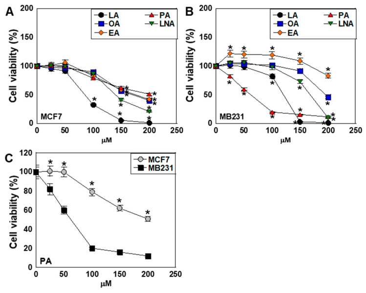
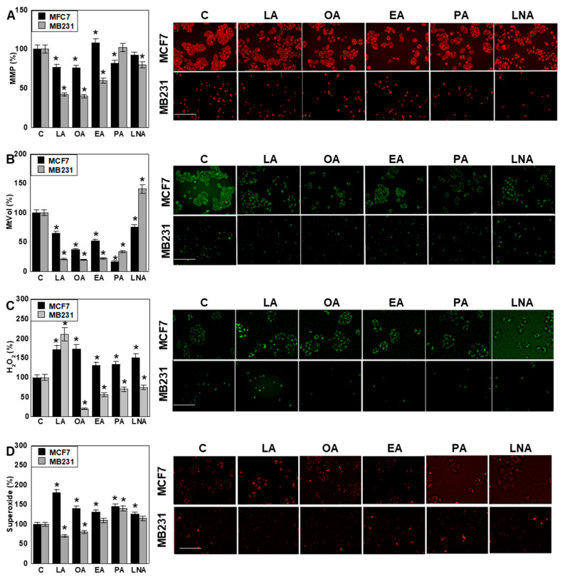
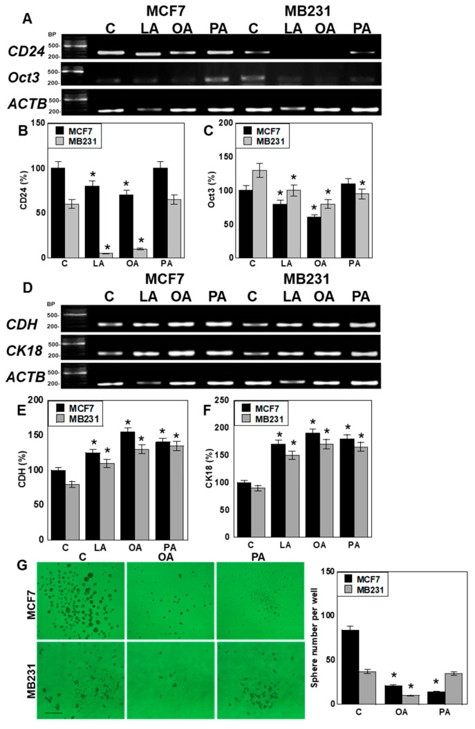
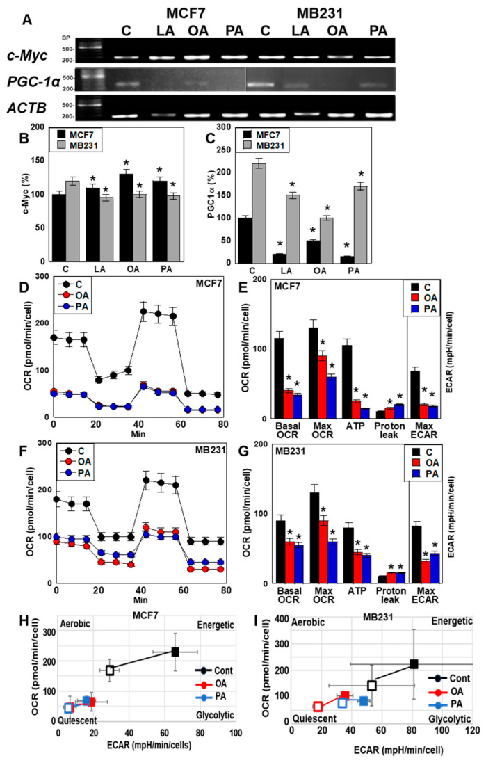
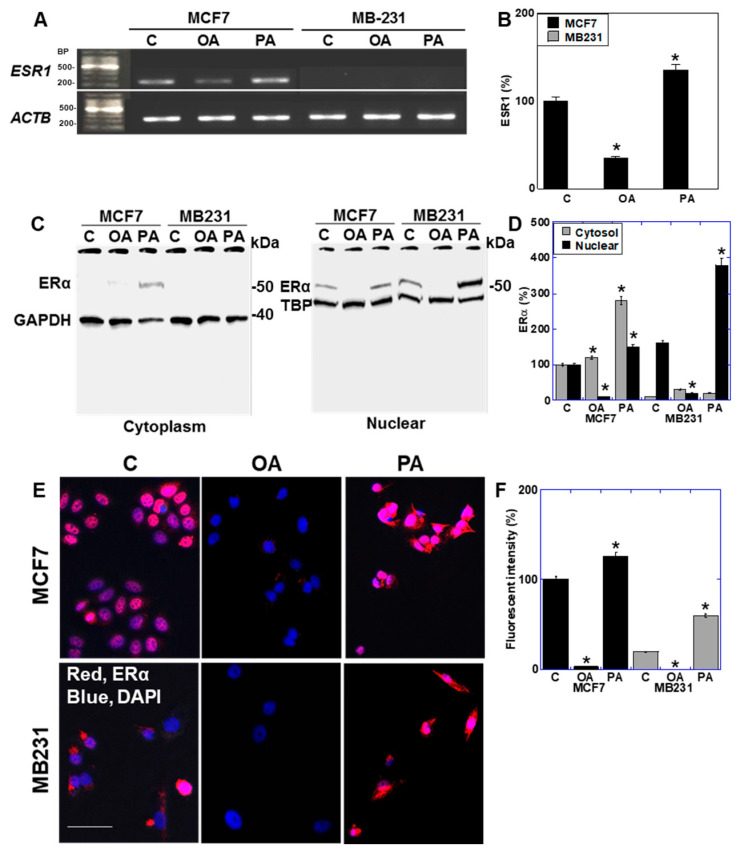
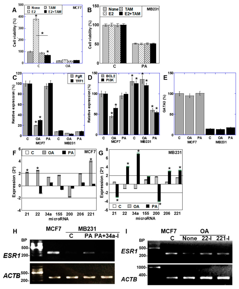
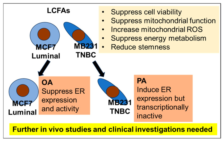
Long-chain fatty acids (LCFAs) have emerged as important regulators of cancer metabolism, but their impact on hormone receptor expression in breast cancer (BCA) remains poorly understood. In this study, we investigated the effects of five LCFAs-linoleic acid (LA), oleic acid (OA), elaidic acid (EA), palmitic acid (PA), and α-linolenic acid (LNA)-on two BCA cell lines: luminal-type MCF7 and triple-negative MDA-MB-231 (MB231). All LCFAs suppressed cell viability and mitochondrial function in a dose-dependent manner, accompanied by decreased membrane potential, increased reactive oxygen species production, and a metabolic shift. Notably, OA reduced both mRNA and nuclear protein levels of estrogen receptor alpha (ERα) in MCF7 cells, leading to impaired responses to estradiol and tamoxifen. In contrast, PA induced nuclear ERα expression in MB231 cells, although ER signaling remained inactive. MicroRNA profiling revealed that OA upregulated ER-suppressive miR-22 and miR-221 in MCF7, while PA increased miR-34a in MB231, contributing to ERα induction. These findings suggest that specific LCFAs modulate ER expression through epigenetic and post-transcriptional mechanisms, altering hormonal responsiveness in BCA. Our results offer new insights into how dietary lipids may influence therapeutic efficacy and tumor behavior by regulating nuclear receptor signaling.

Keywords: breast cancer; estrogen receptor; long-chain fatty acid; triple negative breast cancer.


Conflict of interest statement

The authors declare no conflicts of interest.

Figures

Figure 1
Figure 1
Effect of LCFAs on cell viability of BCA cells. (A,B) The BCA cell lines MCF7 (A) and MT231 (B) were treated with various concentrations of LCFAs for 48 h. (C) Comparison of PA on cell viability between MCF7 and MB231 cells. Cell viability in untreated controls was set at 100%. Error bars: SD from three independent trials. Statistical analysis: ANOVA with Bonferroni correction. * p < 0.05, MCF7 vs. MB231. LCFA, long-chain fatty acid; BCA, breast cancer; LA, linoleic acid; OA, oleic acid; EA, elaidic acid; PA, palmitic acid; LNA, linolenic acid; C, control; ANOVA, analysis of variance; SD, standard deviation.
Figure 2
Figure 2
Effect of LCFAs on mitochondrial function in BCA cells. BCA cells were treated with LCFAs (50 μM) for 48 h. (A) MMP, (B) MtVol, (C) mitochondrial H2O2, and (D) mitochondrial superoxide. Each parameter was expressed relative to the fluorescence intensity of the control, taken as 100%. Scale bar, 50 μm. Error bars: SD from three independent trials. Statistical analysis: ANOVA with Bonferroni correction. * p < 0.05 vs. C. LCFA, long-chain fatty acid; BCA, breast cancer; LA, linoleic acid; OA, oleic acid; EA, elaidic acid; PA, palmitic acid; LNA, linolenic acid; C, control; MMP, mitochondrial membrane potential; MtVol, mitochondrial volume; ANOVA, analysis of variance; SD, standard deviation.
Figure 3
Figure 3
Effect of LCFAs on stemness and differentiation of BCA cells. BCA cells were treated with LCFAs (50 μM) for 48 h. (A) Gene expression of stemness-related genes, namely CD24 and Oct3. (B,C) Semi-quantification of panel A. The MCF7 control was set to 100. (D) Expression of differentiation-related genes, namely CDH and CK18. (E,F) Semi-quantification of panel D. The MCF7 control was set to 100. (G) Sphere formation in the BCA cells. Scale bar, 200 μm. Right panel: Quantification of the left panel. Error bars: SD from three independent trials. Statistical analysis: ANOVA with Bonferroni correction. * p < 0.05 vs. C. LCFA, long-chain fatty acid; BCA, breast cancer; LA, linoleic acid; OA, oleic acid; PA, palmitic acid; C, control; BP, base pair; Oct3, octamer-binding transcription factor 3; ACTB, β-actin; CDH, E-cadherin; CK18, cytokeratin; ANOVA, analysis of variance; SD, standard deviation.
Figure 4
Figure 4
Effect of LCFAs on energy metabolism of BCA cells. BCA cells were treated with LCFAs (50 μM) for 48 h. (A) Gene expression of energy metabolism-related genes, namely c-Myc and PGC-1α. (B,C) Semi-quantification of panels (A,DG). Energy flux analysis of MCF7 (D,F) and MB231 cells (E,G). (H,I) Matrix diagram of energy metabolism. Error bars: SD from three independent trials. Statistical analysis: ANOVA with Bonferroni correction. * p < 0.05 vs. C. LCFA, long-chain fatty acid; BCA, breast cancer; LA, linoleic acid; OA, oleic acid; PA, palmitic acid; C, control; BP, base pair; PGC-1α, peroxisome proliferator-activated receptor gamma coactivator 1-α; ACTB, β-actin; OCR, oxygen consumption rate; ECAR, extracellular acidification rate; Max maximal; C, control; cont, control; ANOVA, analysis of variance; SD, standard deviation.
Figure 5
Figure 5
Effect of LCFAs on expression of estrogen receptor in BCA cells. BCA cells were treated with LCFAs (50 μM) for 48 h. (A) Gene expression of ESR1. (B) Semi-quantification of panel A. (C) Protein levels of ERα in the cytoplasm and nuclei. (D) Semi-quantification of panel D. (E) Immunocytochemistry of ERα. Scale bar, 50 μm. (F) Semi-quantification of the fluorescence intensity in panel E. Error bars: SD from three independent trials. Statistical analysis: ANOVA with Bonferroni correction. * p < 0.05 vs. C.LCFA, long-chain fatty acid; BCA, breast cancer; OA, oleic acid; PA, palmitic acid; C, control; BP, base pair; ESR1, estrogen receptor gene; ER, estrogen receptor; GAPDH, glyceraldehyde-3-phosphate dehydrogenase; TBP, TATA-binding protein; DAPI, 4′,6-diamidino-2-phenylindole; ANOVA, analysis of variance; SD, standard deviation.
Figure 6
Figure 6
Function of ER expressed in BCA cells. BCA cells were treated with LCFAs (50 μM) with or without E2 or TAM for 48 h. (A,B) Effects of E2 and TAM on the viability of MCF7 (A) and MB231 cells (B). (CE) Expression of PgR and TFF1 (C), BCL2 and PI3K (D), and GATA3 (E) based on the data from quantitative PCR. The expression was standardized by ACTB expression. (F,G) Expression of ER-associated miRNAs in MCF7 (F) and MB231 cells (G). (H) ESR1 expression in MB231 cells treated with PA (50 μM) and/or the miR-34a inhibitor (34a-I, 2 μM) for 48 h. (I) ESR1 expression in MCF7 cells treated with OA (50 μM) and/or miR-22 or -221 inhibitors (22-I or 221-I, 2 μM) for 48 h. Error bars: SD from three independent trials. Statistical analysis: ANOVA with Bonferroni correction. * p < 0.05 vs. C. LCFA, long-chain fatty acid; BCA, breast cancer; OA, oleic acid; PA, palmitic acid; C, control; ER, estrogen receptor; E2, estradiol; TAM, tamoxifen; PgR, progesterone receptor; TFF1, trefoil factor-1; BCL2, B-cell CLL/lymphoma 2; PI3K, phosphatidyl inositol-3 kinase; GATA3, GATA-binding protein 3; BP, base pair; ANOVA, analysis of variance; SD, standard deviation.
Figure 7
Figure 7
LCFAs modulate mitochondrial function, energy metabolism, and cellular stemness in breast cancer cells. Notably, OA suppresses ER expression and activity in luminal-type MCF7 cells. In contrast, PA induces ER re-expression in TNBC MB231 cells; however, the receptor remains transcriptionally inactive. Further in vivo studies and clinical validation are necessary to determine the translational relevance of these findings. LCFA, long-chain fatty acids; ROS, reactive oxygen species; TNBC, triple-negative breast cancer; MB231, MDA-MB231; OA, oleic acid; ER, estrogen receptor; PA, palmitic acid.

Similar articles

References

    1. Sung H., Ferlay J., Siegel R.L., Laversanne M., Soerjomataram I., Jemal A., Bray F. Global Cancer Statistics 2020: GLOBOCAN Estimates of Incidence and Mortality Worldwide for 36 Cancers in 185 Countries. CA Cancer J. Clin. 2021;71:209–249. doi: 10.3322/caac.21660. - DOI - PubMed
    1. Perou C.M., Sørlie T., Eisen M.B., van de Rijn M., Jeffrey S.S., Rees C.A., Pollack J.R., Ross D.T., Johnsen H., Akslen L.A., et al. Molecular portraits of human breast tumours. Nature. 2000;406:747–752. doi: 10.1038/35021093. - DOI - PubMed
    1. Reis-Filho J.S., Pusztai L. Gene expression profiling in breast cancer: Classification, prognostication, and prediction. Lancet. 2011;378:1812–1823. doi: 10.1016/S0140-6736(11)61539-0. - DOI - PubMed
    1. Prat A., Cheang M.C., Martín M., Parker J.S., Carrasco E., Caballero R., Tyldesley S., Gelmon K., Bernard P.S., Nielsen T.O., et al. Prognostic significance of progesterone receptor-positive tumor cells within immunohistochemically defined luminal A breast cancer. J. Clin. Oncol. 2013;31:203–209. doi: 10.1200/JCO.2012.43.4134. - DOI - PMC - PubMed
    1. Carey L.A., Berry D.A., Cirrincione C.T., Barry W.T., Pitcher B.N., Harris L.N., Ollila D.W., Krop I.E., Henry N.L., Weckstein D.J., et al. Molecular Heterogeneity and Response to Neoadjuvant Human Epidermal Growth Factor Receptor 2 Targeting in CALGB 40601, a Randomized Phase III Trial of Paclitaxel Plus Trastuzumab With or Without Lapatinib. J. Clin. Oncol. 2016;34:542–549. doi: 10.1200/JCO.2015.62.1268. - DOI - PMC - PubMed

MeSH terms

LinkOut - more resources