. 2025 Jul 14;26(14):6732.
doi: 10.3390/ijms26146732.

The Dichloromethane Fraction of Sanguisorba tenuifolia Inhibits Inflammation in Cells Through Modulation of the p38/ERK/MAPK and NF-κB Signaling Pathway

Affiliations

The Dichloromethane Fraction of Sanguisorba tenuifolia Inhibits Inflammation in Cells Through Modulation of the p38/ERK/MAPK and NF-κB Signaling Pathway

Yue Wang et al. Int J Mol Sci. .

Abstract

Sanguisorba tenuifolia is a wild plant of the genus Sanguisorba officinalis. This study aimed to investigate the regulatory effect of the dichloromethane fraction of Sanguisorba tenuifolia on LPS-induced inflammatory responses in RAW264.7 cells, thereby providing a new scientific basis for the medicinal development of Sanguisorba tenuifolia. Initially, we used 75% ethanol to crudely extract the roots of Sanguisorba tenuifolia, followed by fractional extraction using dichloromethane (CH2Cl2), ethyl acetate (EtOAc), butanol (BuOH), and distilled water (DW) as solvents. By measuring the inhibitory effects of each fractionated extract on NO production, we determined that the SCE (Dichloromethane fraction of Sanguisorba tenuifolia) exhibited the most potent anti-inflammatory activity, leading to its progression to the next experimental stage. Subsequently, we evaluated the effects of SCE on cell viability and LPS-induced inflammatory cytokine secretion in RAW264.7 cells. A rat model of reflux esophagitis was also used to validate the in vivo anti-inflammatory effects of SCE. Additionally, we utilized UPLC/MS-MS to identify and analyze the active components of SCE. The results indicated that SCE could effectively inhibit LPS-induced cellular inflammation by modulating the p38/ERK/MAPK and NF-κB signaling pathways, and also reduced the damage of the esophageal mucosa in rats with reflux esophagitis. UPLC/MS-MS analysis of SCE identified 423 compounds, including 12 active ingredients such as triterpenoids, phenols, and steroids. This discovery not only provides scientific support for the potential of Sanguisorba tenuifolia as an anti-inflammatory agent but also lays the groundwork for the development of new therapeutics for the treatment of inflammatory diseases.

Keywords: MAPK/NF-κB signaling pathway; Sanguisorba tenuifolia; anti-inflammatory; dichloromethane fraction.


Conflict of interest statement

There are no conflicts to declare.

Figures
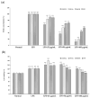
Figure 1
Figure 1
Cells were placed in 96-well plates at a density of 1 × 10⁵ cells/mL. The concentration of the extracts was adjusted to 25 μg/mL, 50 μg/mL, and 100 μg/mL, respectively, and after 1 h of treatment, 1 μg/mL LPS was added and incubated for 18 h. The effect of the graded extracts of Sanguisorba tenuifolia on the amount of NO production for LPS-induced inflammation (a) and on the cell viability (b). Data are expressed as mean ± standard deviation; LPS was statistically analyzed in comparison to normal cells (### p < 0.001), and comparison between each sample and LPS-induced cells (*** p < 0.001,** p < 0.01).
Figure 2
Figure 2
Cells were placed in 96-well plates at a density of 1 × 10⁵ cells/mL. The concentration of extracts was adjusted to 25 μg/mL, 50 μg/mL, and 100 μg/mL, respectively, and after 1 h of treatment, 1 μg/mL LPS was added and incubated for 18 h. The effect of SCE on cell morphology changes.
Figure 3
Figure 3
Determination of TNF-α (a) and IL-1β (b) levels in cell supernatants using ELISA kits. The effects of SCE on the expression levels of iNOS (c) and COX-2 (d) in LPS-induced RAW264.7 cells were detected by immunoblotting. Data are expressed as mean ± standard deviation, and LPS was statistically analyzed in comparison to normal cells (### p < 0.001, ## p < 0.01), and comparison between each sample and LPS-induced cells (*** p < 0.001, ** p < 0.01, * p < 0.05).
Figure 4
Figure 4
Effect of SCE on p38 and ERK phosphorylation levels in LPS-induced RAW264.7 cells detected by immunoblotting. The data are expressed as mean ± standard deviation. LPS was statistically analyzed in comparison to normal cells (### p < 0.001, ## p < 0.01), and comparison between each sample and LPS-induced cells (** p < 0.01, * p < 0.05).
Figure 5
Figure 5
The influence of SCE on the phosphorylation of NF-κB p65 and IκBα (a) and the nuclear translocation of (b) in RAW264.7 cells stimulated by LPS was examined using immunoblotting and immunofluorescence techniques. Data were expressed as mean ± standard deviation. LPS was statistically analyzed in comparison to normal cells (# p < 0.05), and the comparison between each sample and LPS-induced cells (* p < 0.05).
Figure 6
Figure 6
Effects of SCE on esophageal reflux-induced esophageal mucosal damage in rats. Gross (a), the ratio of esophageal damage (b). N, Normal rats; Veh, RE-controlled rats; SCE 100 mg/kg, RE-controlled rats treated with SCE 100 mg/kg; SCE 200 mg/kg, RE-controlled rats treated with SCE 200 mg/kg and R, RE-controlled rats treated with ranitidine 30 mg/kg. ### p < 0.001, *** p < 0.001, vs. RE-controlled rats. Data were expressed as mean ± standard deviation.
Figure 7
Figure 7
Expanded base peak chromatograms from 0 to 25 min obtained by ultra-performance liquid chromatography-tandem mass spectrometry (UPLC-MS/MS) analysis of SCE.
Figure 8
Figure 8
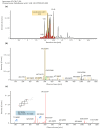
The chromatograph (a), molecular ion peak (b), and fragment ion peak (c) of Ganoderenic acid C in SCE detected by LC-MS/MS (negative ESI source mode) analysis [Color figure can be viewed at wileyonlinelibrary.com].
Figure 9
Figure 9
The chromatograph (a), molecular ion peak (b), and fragment ion peak (c) of Belachinal in SCE detected by LC-MS/MS (negative ESI source mode) analysis [Color figure can be viewed at wileyonlinelibrary.com].

References

    1. Arulselvan P., Fard M.T., Tan W.S., Gothai S., Fakurazi S., Norhaizan M.E., Kumar S.S. Role of Antioxidants and Natural Products in Inflammation. Oxid. Med. Cell. Longev. 2016;2016:5276130. doi: 10.1155/2016/5276130. - DOI - PMC - PubMed
    1. Schromm A.B., Brandenburg K., Loppnow H., Moran A.P., Koch M.H., Rietschel E.T., Seydel U. Biological Activities of Lipopolysaccharides Are Determined by the Shape of Their Lipid A Portion. Eur. J. Biochem. 2000;267:2008–2013. doi: 10.1046/j.1432-1327.2000.01204.x. - DOI - PubMed
    1. Chang X., Zhang T., Wang J., Liu Y., Yan P., Meng Q., Yin Y., Wang S. SIRT5-Related Desuccinylation Modification Contributes to Quercetin-Induced Protection against Heart Failure and High-Glucose-Prompted Cardiomyocytes Injury through Regulation of Mitochondrial Quality Surveillance. Oxid. Med. Cell. Longev. 2021;2021:5876841. doi: 10.1155/2021/5876841. - DOI - PMC - PubMed
    1. Korinek M., Tsai Y.H., El-Shazly M., Lai K.H., Backlund A., Wu S.F., Lai W.C., Wu T.Y., Chen S.L., Wu Y.C., et al. Anti-Allergic Hydroxy Fatty Acids from Typhonium blumei Explored through ChemGPS-NP. Front. Pharmacol. 2017;8:356. doi: 10.3389/fphar.2017.00356. - DOI - PMC - PubMed
    1. Singh N., Baby D., Rajguru J.P., Patil P.B., Thakkannavar S.S., Pujari V.B. Inflammation and Cancer. Ann. Afr. Med. 2019;18:121–126. doi: 10.4103/aam.aam_56_18. - DOI - PMC - PubMed

MeSH terms

LinkOut - more resources