. 2025 Jul 16;26(14):6843.
doi: 10.3390/ijms26146843.

Amine-Modified Diatomaceous Earth Syringe Platform (DeSEI) for Efficient and Cost-Effective EV Isolation

Affiliations

Amine-Modified Diatomaceous Earth Syringe Platform (DeSEI) for Efficient and Cost-Effective EV Isolation

Hyo Joo Lee et al. Int J Mol Sci. .

Abstract

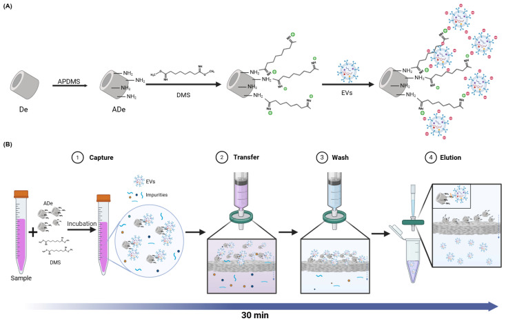
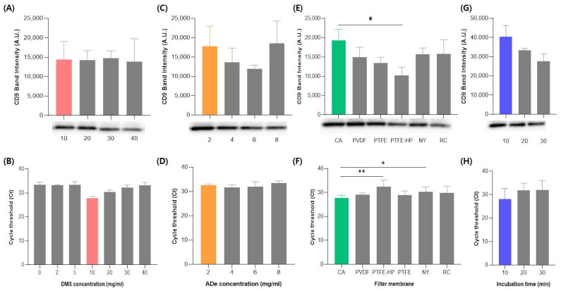
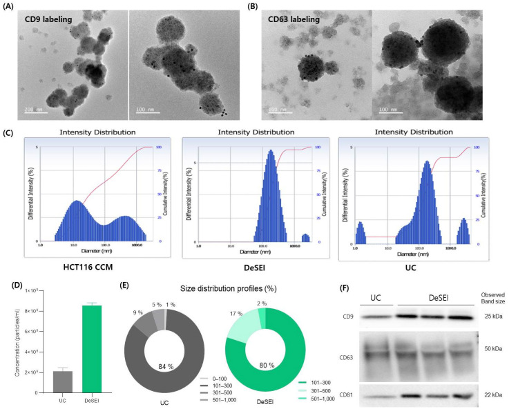
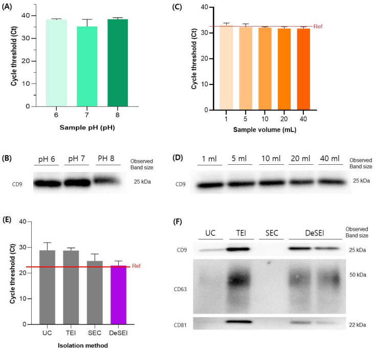
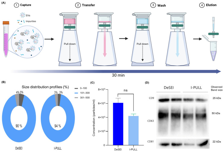
Conventional methods for isolating extracellular vesicles (EVs) are often limited by long processing times, a low purity, and a reliance on specialized equipment. To overcome these challenges, we developed the DeSEI (amine-functionalized Diatomaceous earth-based Syringe platform for EV Isolation), a novel platform employing low-cost, amine-functionalized diatomaceous earth (ADe) within a simple syringe-filter system. The capture mechanism leverages the electrostatic interaction between the positively charged ADe and the negatively charged EV surface, enabling a rapid and efficient isolation. The optimized 30 min protocol yields intact EVs with morphology, size, and protein markers comparable to those from ultracentrifugation, ensuring minimal cellular contamination. Notably, DeSEI exhibited a nearly 60-fold higher recovery efficiency of EV-derived miRNA compared to ultracentrifugation. The platform further proved its versatility with a rapid one-step miRNA extraction protocol and a user-friendly cartridge format. The direct miRNA extraction capability is particularly advantageous for a streamlined biomarker analysis, while the cartridge design illustrates a clear pathway toward developing point-of-care diagnostic tools. The DeSEI offers a promising alternative to existing methods for EV-based research by providing a combination of speed, simplicity, and procedural flexibility that does not require specialized equipment.

Keywords: EV isolation; diatomaceous earth; dimethyl sulfide; extracellular vesicle; nanomaterials; sample preparation.


Conflict of interest statement

Authors Jinkwan Lee and Namheon Kim were employed by the company Infusiontech. The remaining authors declare that the research was conducted in the absence of any commercial or financial relationships that could be construed as a potential conflict of interest.

Figures

Figure 1
Figure 1
A schematic of the principle and the isolation of extracellular endosomes using the DeSEI. (A) A schematic of the capture mechanism. The surface of the DE is functionalized with APDMS to introduce amine groups, creating ADe. DMS then acts as a cross-linker, enabling the capture of negatively charged EVs onto the positively charged ADe surface through electrostatic interactions. (B) The DeSEI workflow. First, ADe and DMS are added to the sample and incubated to facilitate EV capture. Next, the EV-bound ADe is isolated, and debris is removed using a syringe filter. Finally, an elution buffer is used to disrupt the electrostatic bonds, releasing the purified EVs from the ADe. The entire four-step process is completed in under 30 min. (This figure was created with BioRender.com). Abbreviations: DeSEI, amine-functionalized Diatomaceous earth Syringe platform for EV Isolation; ADe, Amine-functionalized Diatomaceous earth; DE, Diatomaceous earth; and EVs, extracellular vesicles.
Figure 2
Figure 2
The optimization of the DeSEI protocol for EV isolation. The EV yield for each parameter was evaluated by quantifying the EV-specific protein marker CD9 via the Western blot analysis and the EV-enriched cargo miRNA-21 via the RT-qPCR. The optimization of the (A,B) DMS concentration, (C,D) ADe concentration, (E,F) syringe filter membrane type, and (G,H) sample incubation time. In all graphs, the selected optimal condition for each parameter is highlighted (DMS in red, ADe in orange, filter type in green, and incubation time in blue). The final optimized protocol was established with a DMS concentration of 10 mg/mL, an ADe concentration of 2 mg/mL, a cellulose acetate (CA) syringe filter, and a 10 min incubation time. Data are presented as mean ± standard deviation (n = 3). Significant differences are indicated as follows: * p < 0.05 and ** p < 0.01. Abbreviations: DeSEI, amine-functionalized Diatomaceous earth Syringe platform for EV Isolation; ADe, Amine-functionalized Diatomaceous earth; DMS, Dimethyl suberimidate dihydrochloride; and EV, extracellular vesicle.
Figure 3
Figure 3
The physicochemical and molecular characterization of EVs isolated by the DeSEI. (A,B) The transmission electron microscopy (TEM) of isolated EVs. Representative images show spherical vesicles of approximately 100–200 nm, confirming their intact morphology. The immunogold labeling demonstrates the presence of the EV surface markers (A) CD9 and (B) CD63, indicated by attached gold nanoparticles (black dots). (C) The dynamic light scattering (DLS) analysis of the particle size distribution. While the initial HCT116 CCM (left) contains a heterogeneous particle population, EVs isolated by the DeSEI (center) show a distinct peak between 100 and 300 nm, a pattern comparable to that of EVs isolated by UC (right). (D,E) The nanoparticle tracking analysis (NTA) of isolated EVs. (D) The particle concentration and (E) size distribution profiles of EVs isolated using UC and DeSEI methods from the same starting sample volume. (F) The Western blot analysis of EV-specific protein markers (tetraspanins CD9, CD63, and CD81) in the DeSEI-isolated EVs. Abbreviations: DeSEI, amine-functionalized Diatomaceous earth Syringe platform for EV Isolation; CCM, cell culture media; UC, ultracentrifugation; and EVs, extracellular vesicles.
Figure 4
Figure 4
The performance and comparative efficiency of the DeSEI. (A,B) The evaluation of the EV isolation efficiency from samples with varying pH levels. (A) RT-qPCR analysis for miRNA-21, where darker green bars correspond to higher pH values (pH 6, 7, and 8). (B) Corresponding Western blot analysis for the CD9 marker. (C,D) A comparison of the EV isolation efficiency across varying sample volumes (1 mL to 40 mL). (C) RT-qPCR analysis for miRNA-21, where darker shades of orange correspond to larger sample volumes. (D) Corresponding Western blot analysis for the CD9 marker. (E,F) The evaluation of the DeSEI performance against other commercial EV isolation methods. (E) RT-qPCR analysis for miRNA-21, where the performance of DeSEI (purple bar) is compared against the other methods (grey bars); the red line indicates the reference value. (F) Corresponding Western blot analysis for the canonical EV markers CD9, CD63, and CD81. Abbreviations: DeSEI, amine-functionalized Diatomaceous earth Syringe platform for EV Isolation; EV, extracellular vesicle; UC, ultracentrifugation; SEC, size exclusion chromatography; and TEI, total exosome isolation kit.
Figure 5
Figure 5
The performance and comparative efficiency of the DeSEI. (A) The workflow of the one-step miRNA extraction using the DeSEI. The initial EV capture and transfer steps are performed as previously described. Subsequently, in the lysis step, a lysis buffer is added, and the sample is incubated for 30 min. A wash step is then performed to remove residual debris. Finally, an elution buffer is added to collect the purified miRNA. The entire five-stage process is completed in under 60 min. (This figure was created with BioRender.com). (B) The optimization of the lysis incubation time, with the optimal duration of 30 min highlighted in orange. (C) The optimization of the additional DMS concentration in the lysis buffer. The highest miRNA yield was highlighted in orange. (D) A comparison of the miRNA extraction efficiency between the integrated one-step DeSEI method and a conventional two-step method, where the DeSEI isolation is performed prior to the miRNA extraction using a commercial kit. Significant differences are indicated as follows: **** p < 0.0001, ns (not significant) p > 0.05. For all optimization and comparison experiments, the extraction efficiency was quantified by measuring miRNA-21 levels via a RT-qPCR. Data are presented as the mean ± standard deviation (n = 3). Abbreviations: DeSEI, amine-functionalized Diatomaceous earth Syringe platform for EV Isolation; EVs, extracellular vesicles.
Figure 6
Figure 6
The schematic and characterization of the I-PULL cartridge with the DeSEI. (A) The I-PULL device for a simplified DeSEI workflow. While the fundamental method is unchanged, the I-PULL cartridge simplifies the process. It features an integrated syringe filter and operates on a vacuum-assisted principle. Loading the sample and pulling down the lower section creates negative pressure, which forces the solution through the filter for rapid separation. (This figure was created with BioRender.com). (B,C) The nanoparticle tracking analysis (NTA) of isolated EVs. (B) Size distribution profiles and (C) the particle concentration of EVs isolated using the DeSEI and I-PULL methods from the same starting sample volume. Significant differences are indicated as follows: ns (not significant) p > 0.05. (D) The Western blot analysis of EV-specific protein markers (tetraspanins CD9, CD63, and CD81) in the DeSEI-isolated and I-PULL-isolated EVs. Abbreviations: DeSEI, amine-functionalized Diatomaceous earth Syringe platform for EV Isolation; EVs, extracellular vesicles.

Similar articles

References

    1. Kalluri R., LeBleu V.S. The biology, function, and biomedical applications of exosomes. Science. 2020;367:6977. doi: 10.1126/science.aau6977. - DOI - PMC - PubMed
    1. Raposo G., Stoorvogel W. Extracellular vesicles: Exosomes, microvesicles, and friends. J. Cell Biol. 2013;200:373–383. doi: 10.1083/jcb.201211138. - DOI - PMC - PubMed
    1. Yanez-Mo M., Siljander P.R., Andreu Z., Zavec A.B., Borras F.E., Buzas E.I., Buzas K., Casal E., Cappello F., Carvalho J., et al. Biological properties of extracellular vesicles and their physiological functions. J. Extracell. Vesicles. 2015;4:27066. doi: 10.3402/jev.v4.27066. - DOI - PMC - PubMed
    1. van Niel G., D’Angelo G., Raposo G. Shedding light on the cell biology of extracellular vesicles. Nat. Rev. Mol. Cell Biol. 2018;19:213–228. doi: 10.1038/nrm.2017.125. - DOI - PubMed
    1. Das S., Lyon C.J., Hu T. A Panorama of Extracellular Vesicle Applications: From Biomarker Detection to Therapeutics. ACS Nano. 2024;18:9784–9797. doi: 10.1021/acsnano.4c00666. - DOI - PMC - PubMed

MeSH terms

LinkOut - more resources