. 2025 Jul 18;26(14):6916.
doi: 10.3390/ijms26146916.

Csn5 Depletion Reverses Mitochondrial Defects in GCN5-Null Saccharomyces cerevisiae

Affiliations

Csn5 Depletion Reverses Mitochondrial Defects in GCN5-Null Saccharomyces cerevisiae

Angela Cirigliano et al. Int J Mol Sci. .

Abstract

In this study, we investigated the mitochondrial defects resulting from the deletion of GCN5, a lysine-acetyltransferase, in the yeast Saccharomyces cerevisiae. Gcn5 serves as the catalytic subunit of the SAGA acetylation complex and functions as an epigenetic regulator, primarily acetylating N-terminal lysine residues on histones H2B and H3 to modulate gene expression. The loss of GCN5 leads to mitochondrial abnormalities, including defects in mitochondrial morphology, a reduced mitochondrial DNA copy number, and defective mitochondrial inheritance due to the depolarization of actin filaments. These defects collectively trigger the activation of the mitophagy pathway. Interestingly, deleting CSN5, which encodes to Csn5/Rri1 (Csn5), the catalytic subunit of the COP9 signalosome complex, rescues the mitochondrial phenotypes observed in the gcn5Δ strain. Furthermore, these defects are suppressed by exogenous ergosterol supplementation, suggesting a link between the rescue effect mediated by CSN5 deletion and the regulatory role of Csn5 in the ergosterol biosynthetic pathway.

Keywords: Saccharomyces cerevisiae; epigenetic regulation; ergosterol; lysine-acetyltransferase; mitochondria; ubiquitin–proteasome pathway.


Conflict of interest statement

The authors declare no conflicts of interest.

Figures

Figure 1
Figure 1
Mitochondrial morphology and movement are altered in gcn5Δ cells. Fluorescence microscopy of wild-type (WT), gcn5Δ, and rho° cells transformed with mtGFP (green), stained with DAPI (blue), and Rhodamine-Phalloidin (red). Fluorescent signals show mitochondria (green), DNA (blue), and the actin cytoskeleton (red). The actin polarization into the buds is indicated by arrows. In gcn5Δ the mitochondrial migration into the bud is reduced. Scale bars: 2 μm.
Figure 2
Figure 2
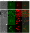
In gcn5Δ, the mitochondrial defects induce the mitophagy detected by fluorescence microscopy. Wild-type (WT), Rapamycin-treated WT, and gcn5Δ and rho° strains have been transformed with GFP-Atg32, which highlights mitochondrial membranes (green), and stained with FM4-64, which stains vacuolar membranes (red). Mitophagy is visible where the green GFP signal is localized within the vacuoles. Scale bars: 2 μm.
Figure 3
Figure 3
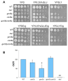
The deletion of CSN5 gene suppresses the mitochondrial phenotypes of gcn5Δ. (A) Growth of serial dilutions of wild-type (WT) and csn5Δ, gcn5Δ, gcn5Δ/csn5Δ, and rho° strains in solid medium containing 2% glucose (fermentation), 0.25% glucose (intermediate), or 3% glycerol (respiration) as a carbon source. Images were taken after three days of growth at 28 °C. (B) Oxygen consumption rate (OCR) of strains grown overnight in 2% glucose-containing medium. (C) Mitochondrial network of strains transformed with mtGFP was observed by fluorescence microscopy (green). Scale bars: 1 μm. (D) qRT-PCR analysis of mtDNA level of the WT and derivative deleted mutants grown in 2% glucose-containing medium. The ratio between nuclear DNA (nDNA) mean value and mtDNA mean value (OXI1/ACT1) was used to overcome the variability among samples caused by total DNA quality. Data derive from at least three independent experiments and statistical significance by Student’s t-test is indicated. ** p < 0.01; * p < 0.05 for mutants versus WT strain.
Figure 4
Figure 4
The deletion of CSN5 suppresses the mitophagy in gcn5Δ. Fluorescence microscopy of wild-type (WT) and csn5Δ, gcn5Δ, and gcn5Δ/csn5Δ strains transformed with GFP-Atg32, which highlights mitochondrial membranes (green), and stained with FM4-64 (vacuolar membranes in red). Scale bars: 2 μm.
Figure 5
Figure 5
Ergosterol content in mutant strains. The esterified ergosterol was measured by Hig- Performance Liquid Chromatography (HPLC) analysis in wild-type (WT) and rho°, gcn5Δ, csn5Δ, and gcn5Δ/csn5Δ cells. Values were reported as ergosterol percentage in the mutants compared to that of wild-type cells. Data derive from at least three independent experiments and statistical significance by Student’s t-test is indicated. ** p < 0.01; for mutants versus WT strain.
Figure 6
Figure 6
Respiration phenotypes of the mutant strains treated with ergosterol. (A) Growth of wild-type (WT), rho°, gcn5Δ, csn5Δ, and gcn5Δ/csn5Δ mutants in a solid medium (serial dilutions) containing 2% glucose, 0.25% glucose, or 3% glycerol as a carbon source (upper panels) and on the same media supplemented with 0.02 mg/mL ergosterol. All strains were grown overnight in 2% glucose-liquid-containing medium with the addition of 0.02 mg/mL ergosterol before being plated on solid media. Images were taken after three days of growth at 28 °C. (B) Oxygen consumption rate (OCR) of indicated strains grown in 2% glucose-containing medium with the addition of 0.02 mg/mL ergosterol. Data derive from at least three independent experiments and statistical significance by Student’s t-test is indicated. ** p < 0.01; * p < 0.05 for mutants versus WT strain.
Figure 7
Figure 7
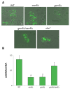
Ergosterol supplementation suppresses the mitochondrial morphology defects and the mtDNA instability. (A) Fluorescence microscopy of wild-type (WT) and rho°, gcn5Δ, csn5Δ, and gcn5Δ/csn5Δ mutants grown in 2% glucose medium with the addition of 0.02 mg/mL ergosterol. The strains were transformed with mtGFP to visualize the mitochondrial network (green). Scale bars: 1 μm. (B) qRT-PCR analysis of mtDNA level of the wild-type and the mutants grown as in (A). The ratio between nuclear DNA (nDNA) mean value and mtDNA mean value (OXI1/ACT1) was used to overcome the variability among samples caused by total DNA quality. Data derive from at least three independent experiments and statistical significance by Student’s t-test is indicated. * p < 0.05 for deleted versus WT strain.

Similar articles

References

    1. Soffers J.H.M., Workman J.L. The SAGA Chromatin-Modifying Complex: The Sum of Its Parts Is Greater than the Whole. Genes Dev. 2020;34:1287–1303. doi: 10.1101/gad.341156.120. - DOI - PMC - PubMed
    1. Chou K.Y., Lee J.-Y., Kim K.-B., Kim E., Lee H.-S., Ryu H.-Y. Histone Modification in Saccharomyces cerevisiae: A Review of the Current Status. Comput. Struct. Biotechnol. J. 2023;21:1843–1850. doi: 10.1016/j.csbj.2023.02.037. - DOI - PMC - PubMed
    1. Patel J.H., Du Y., Ard P.G., Phillips C., Carella B., Chen C.-J., Rakowski C., Chatterjee C., Lieberman P.M., Lane W.S., et al. The C-MYC Oncoprotein Is a Substrate of the Acetyltransferases hGCN5/PCAF and TIP60. Mol. Cell. Biol. 2004;24:10826–10834. doi: 10.1128/MCB.24.24.10826-10834.2004. - DOI - PMC - PubMed
    1. Canzonetta C., Leo M., Guarino S.R., Montanari A., Francisci S., Filetici P. SAGA Complex and Gcn5 Are Necessary for Respiration in Budding Yeast. Biochim. Biophys. Acta (BBA)—Mol. Cell Res. 2016;1863:3160–3168. doi: 10.1016/j.bbamcr.2016.10.002. - DOI - PubMed
    1. Montanari A., Leo M., De Luca V., Filetici P., Francisci S. Gcn5 Histone Acetyltransferase Is Present in the Mitoplasts. Biol. Open. 2019;8:bio041244. doi: 10.1242/bio.041244. - DOI - PMC - PubMed

MeSH terms

Substances

LinkOut - more resources