. 2025 Jul 20;26(14):6971.
doi: 10.3390/ijms26146971.

Design of a Multi-Epitope Vaccine Based on Fasciola gigantica Cathepsin B and Evaluation of Immunological Responses in Mice

Affiliations

Design of a Multi-Epitope Vaccine Based on Fasciola gigantica Cathepsin B and Evaluation of Immunological Responses in Mice

Supanan Chansap et al. Int J Mol Sci. .

Abstract

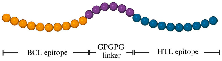
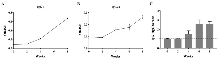
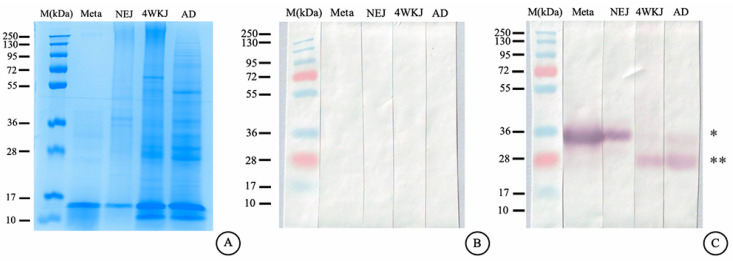
Fasciola gigantica (F. gigantica) is a vital parasite that causes fasciolosis. Liver fluke infections affect livestock animals, and the Fasciola species (Fasciola spp.) vaccine has been tested for many types of these diseases. Currently, computer-based vaccine design represents an attractive alternative for constructing vaccines. Thus, this study aimed to design the epitopes of linear B-cells (BCL) and helper T lymphocytes (HTL) using an immunoinformatic approach and to investigate in silico and the mice's immune response. A non-conserved host region, overlapping F. gigantica cathepsin B proteins (FgCatB), and the highest conserved residue percentages were the criteria used to construct epitopes. The GPGPG linker was used to link epitopes in the multi-epitope Fasciola gigantica cathepsin B (MeFgCatB) peptide. The MeFgCatB peptide has high antigenicity, non-allergenicity, non-toxicity, good solubility, and a high-quality structure. The molecular docking between the MeFgCatB peptide and Toll-like receptor 2 (TLR-2) was evaluated. The IgM, IgG1, and IgG2 levels were elevated in silico. In mice, the MeFgCatB peptide was synthesized and administered as an injection. The MeFgCatB-specific IgG1 and IgG2a levels were elevated after week 2, showing a predominance of IgG1. The rFgCatB1, rFgCatB2, and rFgCatB3 were detected using the MeFgCatB peptide-immunized sera. The MeFgCatB peptide-immunized sera were detected at approximately 28-34 kDa in the whole body. In addition, the MeFgCatB immunized sera can positively signal at the caecal epithelium in the NEJ, 4WKJ, and adult stages. In summary, the MeFgCatB peptide is able to induce mixed Th1/Th2 immune responses with Th2 dominating and to detect the native protein of F. gigantica. The MeFgCatB peptide should help against F. gigantica in future experiments.

Keywords: Cathepsin B; Fasciola gigantica; fasciolosis; immunoinformatic; multi-epitope; vaccines.


Conflict of interest statement

The authors declare no conflicts of interest.

Figures

Figure 1
Figure 1
BCL epitopes of the F. gigantica sequences. (A) FgCatB1, (B) FgCatB2, and (C) FgCatB3 are linear B-cell epitopes. The yellow highlighted regions above the threshold line are BCL epitopes. The green highlighted regions below the threshold line are non-BCL epitopes. The cut-off score is 0.5 (where the Y and X axes represent the scores and positions of the residues of the sequence, respectively).
Figure 2
Figure 2
The BCL and HTL epitope sequences were selected and constructed. The mature protein is indicated by a solid arrow, while the pro-region is indicated by an open arrow. The region of the conserved host is indicated by a yellow label. The group of overlapped BCL and HTL epitopes is denoted by the cyan and green labels, respectively. The novel HTL and BCL epitope sequences are denoted by red and blue letters, respectively. The conserved residue locations of the FgCatBs are shown by an asterix (*), and the conservation across amino acid groups of similar FgCatBs features is indicated by a colon (:). The conservation between amino acid groups of the weakly comparable FgCatBs characteristics is indicated by the period (.).
Figure 3
Figure 3
Schematic representation of the final MeFgCatB peptide. The length of the peptide is 28 aa. The yellow, violet, and blue colors represent the novel BCL epitope, the GPGPG linker, and the novel HTL epitope.
Figure 4
Figure 4
MeFgCatB peptide sequence identity matrix. Sequence identity matrix showing pairwise identity (0–100%) between the MeFgCatB peptide sequence and mature cathepsin B sequences from both the host and F. gigantica. MeFgCatB, multi-epitope-based F. gigantica cathepsin B; FgCatB1, F. gigantica cathepsin B1; FgCatB2, F. gigantica cathepsin B2; FgCatB3, F. gigantica cathepsin B3; MmCatB, Mus musculus cathepsin B; HsCatB, Homo sapiens cathepsin B; BtCatB, Bos taurus cathepsin B; ChCatB, Capra hircus cathepsin B.
Figure 5
Figure 5
Prediction, refinement, and validation of the MeFgCatB peptide tertiary structure. (A) The MeFgCatB peptide’s tertiary structural alignment was indicated in the refined (cyan) and unrefined (green) models. (B) The Z-score (black dot) represents the model’s overall quality. Distinct structural groups have distinct colors (dark blue and light blue, respectively) depending on the source (X-ray, NMR). The calculated Z-score was −1.37. (C) Ramachandran plot of the unrefined model shows 26.7% in the most favored region, 53.3% in the additional allowed region, and 31% in the generously allowed region. (D) The refined model’s Ramachandran plot displays 100% in the most favored region. Red, dark yellow, and light-yellow colors represent the most favored region, the additional allowed region, and the generously allowed region, respectively.
Figure 6
Figure 6
Predicting discontinuous and continuous B-cell epitopes. The discontinuous B-cell epitopes are shown in (A,B). The continuous B-cell epitopes are shown in (C,D). Cyan and yellow colors represent the epitope residues and the rest of the sequence, respectively.
Figure 7
Figure 7
MeFgCatB peptide with TLR-2 molecular docking. A green stick and surface were used to identify the TLR-2 receptor. The molecular interaction was marked with red dotted lines. Magenta, orange, and yellow colors indicate ASN1-PHE266, ARG14-TYP376, and ARG7-SER346 residue interaction, respectively. The rest of the MeFgCatB peptide sequence indicates a cyan color.
Figure 8
Figure 8
MD simulation analysis: (A) The MeFgCatB peptide–TLR-2 complex. NMA areas represent low mobility (red arrow) to high mobility (blue arrow). (B) The B-factor is the RMS and regional mobility score from 0 (lowest) to 1 (highest). (C) The deformability graph of the individual residues in the complex with lower distortion. (D) The eigenvalue is the motion stiffness score as 5.241091 × 10⁻⁶. (E) The eigenvalues, which show the individual (purple) and cumulative (green) variance, have a correlation that is inverse to the variance. (F) Correlated (red), uncorrelated (blue), and anti-correlated (white) motions are indicated by the covariance, which is the interaction between pairs of residues. (G) The elastic network model is a connection between two atoms. The grey indicates a higher protein stiffness in regions.
Figure 9
Figure 9
In silico immune response simulation: (A) antibody and antigen levels; (B) cytokine concentrations; (C) B-cell population by entity state; (D) T-cell population by entity state; (E) macrophage (MA) population per state; (F) dendritic cell (DC) population per state.
Figure 10
Figure 10
Levels of MeFgCatB-specific IgG1 and IgG2a antibodies: (A) the levels of MeFgCatB-specific IgG1 antibodies; (B) the levels of MeFgCatB-specific IgG2a antibodies; (C) calculated IgG1/IgG2a ratios for each mouse. The dashed line represents the IgG1/IgG2a ratio that equals 1.
Figure 11
Figure 11
The MeFgCatB-specific IgG1 levels against rFgCatB1, rFgCatB2, rFgCatB3, and MeFgCatB (A), and the MeFgCatB-specific IgG2a levels against rFgCatB1, rFgCatB2, rFgCatB3, and MeFgCatB (B).
Figure 12
Figure 12
The Coomassie-stained SDS-PAGE of the WB’s F. gigantica and immunoblotting analysis. (A) Coomassie-stained SDS-PAGE showing protein profiles of whole-body extracts from metacercariae (Meta), newly excysted juveniles (NEJ), 4-week juveniles (4WKJ), and adults (AD). (B) The pre-immunized mice serum did not show a positive band in the WB’s F.gigantica in all stages (Meta lane–AD lane). (C) The MeFgCatB immunized mice serum detected a positive band in the WB’s F.gigantica in all stages (Meta lane–AD lane). M, marker lane; Meta, metacercariae; NEJ, newly excysted juvenile; 4WKJ, 4 weeks juvenile; AD, adult. Asterix (*) indicates a molecular weight of approximately 34 kDa. Double asterisks (**) indicate a molecular weight of approximately 28 kDa.
Figure 13
Figure 13
The MeFgCatB peptide-immunized sera localization. (A,C,E) Pre-immunized sera that were not stained in the NEJ, 4WKJ, and adult stages of the F. gigantica section were utilized as the negative control. (B,D,F) Purple staining in the cecal epithelium (Ca) of the NEJ, 4WKJ, and adult stages of F. gigantica tissue indicated the presence of MeFgCatB peptide-immunized sera. The parenchyma (Pc), vitelline gland (Vi), and tegumental cell (Tg) were not stained.
Figure 14
Figure 14
Mice immunization protocol schematic. The MeFgCatB peptide with Quil-A adjuvant was administered to mice three times, two weeks apart (prime, first boost, and second boost). Blood was collected at the time point. All mice were terminated at week 8 for further analysis.

Similar articles

References

    1. Nyindo M., Lukambagire A.H. Fascioliasis: An Ongoing Zoonotic Trematode Infection. BioMed Res. Int. 2015;1:1–8. doi: 10.1155/2015/786195. - DOI - PMC - PubMed
    1. Mostafa W., Abdel-Rady A., El-Dakroury M.F., Felefel W. Field Trials to Evaluate Five Fasciolicides against Natural Liver Fluke Infection in Cattle and Sheep in Egypt. Int. J. Vet. Sci. 2023;12:76–81. doi: 10.47278/journal.ijvs/2022.160. - DOI
    1. Tolan R.W. Fascioliasis Due to Fasciola hepatica and Fasciola gigantica Infection: An Update on This “neglected” Neglected Tropical Disease. Lab. Med. 2011;42:107–116. doi: 10.1309/LMLFBB8PW4SA0YJI. - DOI
    1. Rosas-Hostos Infantes L.R., Paredes Yataco G.A., Ortiz-Martínez Y., Mayer T., Terashima A., Franco-Paredes C., Gonzalez-Diaz E., Rodriguez-Morales A.J., Bonilla-Aldana D.K., Vargas Barahona L., et al. The Global Prevalence of Human Fascioliasis: A Systematic Review and Meta-Analysis. Ther. Adv. Infect. Dis. 2023;10:20499361231185413. doi: 10.1177/20499361231185413. - DOI - PMC - PubMed
    1. Winaya I.B.O., Oka I.B.M., Adnyana I.B.W., Sudipa P.H. Fibrosis and Collagen-I Accumulation in Bali Cattle Liver Tissue Infected with Fasciola gigantica. Int. J. Vet. Sci. 2023;12:224–229. doi: 10.47278/journal.ijvs/2022.179. - DOI

LinkOut - more resources