. 2025 Jul 21;26(14):7004.
doi: 10.3390/ijms26147004.

Titin's Intrinsically Disordered PEVK Domain Modulates Actin Polymerization

Affiliations

Titin's Intrinsically Disordered PEVK Domain Modulates Actin Polymerization

Áron Gellért Altorjay et al. Int J Mol Sci. .

Abstract

The multi-domain muscle protein titin provides elasticity and mechanosensing functions to the sarcomere. Titin's PEVK domain is intrinsically disordered due to the presence of a large number of prolines and highly charged residues. Although PEVK does not have canonical actin-binding motifs, it has been shown to bind F-actin. Here, we explored whether the PEVK domain may also affect actin assembly. We cloned the middle, 733-residue-long segment (called PEVKII) of the full-length PEVK domain, expressed in E. coli and purified by using His- and Avi-tags engineered to the N- and C-termini, respectively. Actin assembly was monitored by the pyrene assay in the presence of varying PEVKII concentrations. The structural features of PEVKII-associated F-actin were studied with atomic force microscopy. The added PEVKII enhanced the initial and log-phase rates of actin assembly and the peak F-actin quantity in a concentration-dependent way. However, the critical concentration of actin polymerization was unaltered. Thus, PEVK accelerates actin polymerization by facilitating its nucleation. This effect was highlighted in the AFM images of F-actin-PEVKII adsorbed to the supported lipid bilayer. The sample was dominated by radially symmetric complexes of short actin filaments. PEVK's actin polymerization-modulating effect may, in principle, have a function in regulating sarcomeric actin length and turnover. Altogether, titin's PEVK domain is not only a non-canonical actin-binding protein that regulates sarcomeric shortening, but one that may modulate actin polymerization as well.

Keywords: AlphaFold; PEVK; actin paracrystal; atomic force microscopy; intrinsically unstructured protein domain; polymerization assay; pyrene actin; supported lipid bilayer; titin.


Conflict of interest statement

The authors declare no conflicts of interest.

Figures

Figure 1
Figure 1
(a) Amino acid sequence of PEVKII. Non-PEVKII amino acids are underlined. The N-terminal His6-tag and the C-terminal Avi-tag sequences are shown in bold. The thrombin recognition site is indicated in italics. (b) SDS-PAGE of the proteins used in the experiments. Lane 1, molecular weight markers; lanes 4–8, bovine serum albumin (BSA) in increasing amounts (50, 100, 250, 500, 1000 µg), used for calibrating total protein mass; lane 10, cloned, expressed, and purified PEVKII segment. Note that even though the expected molecular weight of PEVKII is ~80 kDa, its band is positioned above 130 kDa because it is highly charged (net charge −64); hence, its electrophoretic motion is retarded.
Figure 2
Figure 2
Spectroscopic analysis of pyrene-labeled actin polymerized in the presence of PEVKII. (a) Fluorescence emission spectra (λex 365 nm) recorded after 800 s of actin polymerization. (b) Fluorescence excitation spectra (λem 407 nm) recorded after 800 s of actin polymerization. (c) Time-dependent changes in fluorescence emission, measured at 407 nm (λex 365 nm) across a time span of 800 s. Actin concentration was 4 µM, and the ratio of pyrene-labeled actin was 10%. PEVKII concentration was 5.5 nM. Green dotted and red continuous lines indicate control (actin only) and PEVKII-dependent traces, respectively.
Figure 3
Figure 3
Effect of PEVKII on actin polymerization (4 µM actin, 10% pyrene-labeled). (a) Time trace of fluorescence emission (λex 365 nm, λem 407 nm) across a span of 2000 s. (b) Zoomed-in part of the trace in the time window of 0–40 s, which reflects the initial assembly rates. (c) Zoomed-in part of the trace in the time window of 70–130 s, which accommodates the region in the half-maximal fluorescence emission intensity. (d) Initial rate of actin polymerization as a function of PEVKII concentration. Error bars correspond to the error of linear fit. (e) Half-maximal actin polymerization rate as a function of PEVKII concentration. Error bars correspond to the error of linear fit. (f) Peak F-actin quantity as a function of PEVKII concentration. (g) Time to peak fluorescence emission intensity, as a function of PEVKII concentration. Inset shows the same function on the logarithmic scale.
Figure 4
Figure 4
Effect of PEVKII on ongoing actin polymerization (4 µM actin, 10% pyrene-labeled). PEVKII was added to the same final concentration (125 nM), but at different times of the polymerization process: at the start (0 s, green segmented line), after 500 s (red dotted line), and after 1500 s (blue continuous line). When PEVKII was added after 500 s, the rate of actin polymerization increased suddenly 3.4-fold. By contrast, when added after 1500 s, actin polymerization rate slightly decreased (0.67-fold).
Figure 5
Figure 5
Effect of actin concentration on the PEVKII-induced changes. (a) Time trace of fluorescence emission (λec 365 nm, λem 407 nm) across a span of 2000 s in the presence of 16.25 nM PEVKII and different concentrations of actin (0.4–4.0 µM, 10% pyrene labeled). Fluorescence intensity was measured under continuous illumination. (b) Time trace of fluorescence emission across a span of 8000 s in the presence of 60 nM PEVKII and different concentrations of actin (0.05–2.0 µM, 10% pyrene labeled). The sample was illuminated only during the intermittent (every 10 min) spectroscopic measurement. (c) Fluorescence emission intensity as a function of actin concentration in steady-state assembly conditions (after 8000 s of actin polymerization).
Figure 6
Figure 6
Effect of PEVK on actin filament structure and organization, measured with AFM. (a) Height-contrast AFM image of control actin filaments forming paracrystal on a supported lipid bilayer (SLB). A quantity of 4 µM 10% labeled pyrene actin was polymerized for 1000 s and pipetted on the SLB (DPPC:DPEPC, 1:1 molar ratio). (b) Two-dimensional fast Fourier transform (2D FFT) of the control F-actin AFM image. Inset, amplitude (A) versus spatial frequency (SF) plot along the direction indicated by the white arrowhead in the FFT. The peaks in the spectrum indicate periodicities ranging between 13.3 nm and 200 nm (corresponding to SF values between 75 × 106–5 × 10⁶ m−1). (c,e,g) Height-contrast AFM images of actin filaments polymerized in the presence of PEVKII (11 nM). Light-blue asterisk indicates a focal point from which filaments radiate. Light-blue arrowheads point at granular particles that likely correspond to PEVKII molecules. (d,f,h) Two-dimensional FFT images of the corresponding F-actin–PEVKII AFM images.
Figure 7
Figure 7
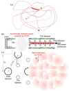
(a) Structure of PEVKII predicted by AlphaFold3. An essentially completely disordered structure is predicted, revealed by low confidence scores (98% of residues possess pLDDT scores lower than 70). The short alpha helical region corresponds to the stretch of the peptide between Asp73 and Glu90. (b) Schematic model of how the intrinsically unstructured PEVK may interact with actin in the sarcomere. The oval indicates the approximate region explored by the PEVK domain. (c) Schematics of the cross-sectional lattice in the overlap region (left) and the I-band (right) of the sarcomere. d10 is the thick-filament lattice spacing (37.4 nm) in the 1.0 x-ray-crystallographic plane, and RG is the radius of gyration (11.7 nm) of the full-length PEVK domain. The two dotted lines correspond to the axes, projected in the transverse sarcomeric plane, of titin molecules interconnecting the tip of the thick filament and the surface of the thin filament (reached at ~100 nm from the Z-line), assuming that each of the six titins running along the thick-filament surface binds to a different thin filament.

Similar articles

References

    1. Wang K., McClure J., Tu A. Titin: Major myofibrillar components of striated muscle. Proc. Natl. Acad. Sci. USA. 1979;76:3698–3702. doi: 10.1073/pnas.76.8.3698. - DOI - PMC - PubMed
    1. Maruyama K. Connectin, an elastic protein from myofibrils. J. Biochem. 1976;80:405–407. doi: 10.1093/oxfordjournals.jbchem.a131291. - DOI - PubMed
    1. Granzier H.L., Labeit S. Discovery of Titin and Its Role in Heart Function and Disease. Circ. Res. 2025;136:135–157. doi: 10.1161/CIRCRESAHA.124.323051. - DOI - PubMed
    1. Dutta D., Nguyen V., Campbell K.S., Padron R., Craig R. Cryo-EM structure of the human cardiac myosin filament. Nature. 2023;623:853–862. doi: 10.1038/s41586-023-06691-4. - DOI - PMC - PubMed
    1. Tamborrini D., Wang Z., Wagner T., Tacke S., Stabrin M., Grange M., Kho A.L., Rees M., Bennett P., Gautel M., et al. Structure of the native myosin filament in the relaxed cardiac sarcomere. Nature. 2023;623:863–871. doi: 10.1038/s41586-023-06690-5. - DOI - PMC - PubMed

LinkOut - more resources