. 2025 Jul 14;16(7):822.
doi: 10.3390/genes16070822.

Integrating Imaging and Genomics in Amelogenesis Imperfecta: A Novel Diagnostic Approach

Affiliations

Integrating Imaging and Genomics in Amelogenesis Imperfecta: A Novel Diagnostic Approach

Tina Leban et al. Genes (Basel). .

Abstract

Background/Objectives: Amelogenesis imperfecta (AI) represents a heterogeneous group of inherited disorders affecting the quality and quantity of dental enamel, making clinical diagnosis challenging. This study aimed to identify genetic variants in Slovenian patients with non-syndromic AI and to evaluate enamel morphology using radiographic parameters. Methods: Whole exome sequencing (WES) was performed on 24 AI patients and their families. Panoramic radiographs (OPTs) were analyzed using Fiji ImageJ to assess crown dimensions, enamel angle (EA), dentine angle (DA), and enamel-dentine mineralization ratio (EDMR) in lower second molar buds, compared to matched controls (n = 24). Two observers independently assessed measurements, and non-parametric tests compared EA, DA, and EDMR in patients with and without disease-causing variants (DCVs). Statistical models, including bootstrap-validated random forest and logistic regression, assessed variable influences. Results: DCVs were identified in ENAM (40% of families), AMELX (15%), and MMP20 (10%), including four novel variants. AI patients showed significant enamel deviations with high reproducibility, particularly in hypomineralized and hypoplastic regions. DA and EDMR showed significant correlations with DCVs (p < 0.01). A bootstrap-validated random forest model yielded a 90% (84.0-98.0%) AUC-estimated predictive power. Conclusions: These findings highlight a novel and reproducible radiographic approach for detecting developmental enamel defects in AI and support its diagnostic potential.

Keywords: amelogenesis imperfecta; disease-causing variants; imaging genomics; molecular genetics; panoramic; radiography.


Conflict of interest statement

The authors declare no conflicts of interest. The funders had no role in the design of the study; in the collection, analyses, or interpretation of data; in the writing of the manuscript; or in the decision to publish the results.

Figures

Figure 1
Figure 1
(A) The illustration shows the implementation of the enamel angle (EA) and the dentine angle (DA) determined on lower permanent second molar buds, as observed in a panoramic radiograph. (B) To assess the relationship between enamel and dentine mineralization, the mean grey values for enamel and dentine (EDMR) are calculated from the middle third of the red-drawn line passing through the enamel and the middle third of the line passing through the dentine, respectively.
Figure 2
Figure 2
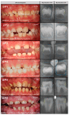
Dental phenotypes (left) and radiographic evaluation of tooth buds 37 and 47 (right) are shown for each of the 24 AI patients from 20 families. The patient number and family number are shown in the upper left corner of the clinical dental image, while the patient’s age and gender are indicated in the upper right corner. The identified genetic variant is listed in the lower left corner; a slash (“/”) denotes cases in which no convincing genetic variant was identified. The colored lines schematically indicate the measured enamel and dentin angles. Note that the angles were measured on both the mesial and distal sides of each tooth bud.
Figure 2
Figure 2
Dental phenotypes (left) and radiographic evaluation of tooth buds 37 and 47 (right) are shown for each of the 24 AI patients from 20 families. The patient number and family number are shown in the upper left corner of the clinical dental image, while the patient’s age and gender are indicated in the upper right corner. The identified genetic variant is listed in the lower left corner; a slash (“/”) denotes cases in which no convincing genetic variant was identified. The colored lines schematically indicate the measured enamel and dentin angles. Note that the angles were measured on both the mesial and distal sides of each tooth bud.
Figure 3
Figure 3
(A) Variable importance in predicting the presence of disease-causing variants (DCVs), as determined by two models: random forest (RF, marked in blue) and logistic regression model (LRM, marked in red). The RF model highlights the influence of enamel angle (EA), dentine angle (DA), and the enamel–dentine mineralization ratio (EDMR) on DCV prediction. Width (W), height (H), and their ratio show minimal impact. In contrast, the LRM identifies DA as the most influential predictor, with a moderate contribution from EDMR. The distributions of W, H, and their ratio are long and shallow, likely due to high inter-correlation among these variables. (B) Predictive performance of both models across 50 iterations of training and validation, evaluated using the area under the curve (AUC). The RF model (blue) shows a median AUC of 90.0% with an interquartile range (IQR) of 84.0–94.0%, while the LRM (red) shows a median AUC of 84.0% with an IQR of 78.0–90.0%.
Figure 4
Figure 4
The relationship between (A) mean EA and EDMR and (B) mean DA and EDMR in AI patients (n = 24) and the control group (n = 24). Clin.(+)/Gen.(+) refers to affected individuals with identified variants in AI-associated genes. Each dot represents an individual carrying a variant in one of the following genes: red (ENAM), brown (AMELX), orange (MMP20), purple (FAM83H), and blue (LAMB3). Clin.(+)/Gen.(−) represents affected individuals in whom no convincing pathogenic variant was found in AI-associated genes. Green dots represent unaffected individuals in the control group. The yellow area represents the hypomineralization region, while the red area represents the hypoplastic region. Cut-off values for EA, DA, and EDMR, determined based on the results from healthy teeth in the control group, are 14.63°, 7.22°, and 1.06, respectively.

Similar articles

References

    1. Witkop C.J. Amelogenesis imperfecta, dentinogenesis imperfecta and dentin dysplasia revisited: Problems in classification. J. Oral Pathol. 1988;17:547–553. doi: 10.1111/j.1600-0714.1988.tb01332.x. - DOI - PubMed
    1. Crawford P.J., Aldred M., Bloch-Zupan A. Amelogenesis imperfecta. Orphanet J. Rare Dis. 2007;2:17. doi: 10.1186/1750-1172-2-17. - DOI - PMC - PubMed
    1. Smith C.E.L., Poulter J.A., Antanaviciute A., Kirkham J., Brookes S.J., Inglehearn C.F., Mighell A.J. Amelogenesis imperfecta: Genes, Proteins, and Pathways. Front. Physiol. 2017;8:435. doi: 10.3389/fphys.2017.00435. - DOI - PMC - PubMed
    1. Nikolopoulos G., Smith C.E.L., Poulter J.A., Murillo G., Silva S., Lamb T., Berry I.R., Brown C.J., Day P.F., Soldani F., et al. Spectrum of pathogenic variants and founder effects in amelogenesis imperfecta associated with MMP20. Hum. Mutat. 2021;42:567–576. doi: 10.1002/humu.24187. - DOI - PubMed
    1. Pavlic A., Petelin M., Battelino T. Phenotype and enamel ultrastructure characteristics in patients with ENAM gene mutations g.13185-13186insAG and 8344delG. Arch. Oral Biol. 2007;52:209–217. doi: 10.1016/j.archoralbio.2006.10.010. - DOI - PubMed

LinkOut - more resources