Review
. 2025 Jul 10;14(14):4902.
doi: 10.3390/jcm14144902.

Screening and Procedural Guidance for Mitral Transcatheter Edge-to-Edge Repair (M-TEER)

Affiliations
Review

Screening and Procedural Guidance for Mitral Transcatheter Edge-to-Edge Repair (M-TEER)

Andromahi Zygouri et al. J Clin Med. .

Abstract

Mitral regurgitation (MR) is a common valvular heart disease associated with significant morbidity and mortality. For patients at high or prohibitive surgical risk, mitral transcatheter edge-to-edge repair (M-TEER) offers a less invasive alternative to surgery. This review outlines key aspects of patient selection and procedural planning for M-TEER, with a focus on clinical and echocardiographic criteria essential for success. Comprehensive imaging-especially 2D and 3D transesophageal echocardiography-is critical to assess leaflet anatomy, coaptation geometry, and mitral valve area. Selection criteria differ between primary and secondary MR and are guided by trials such as COAPT and MITRA-FR. Optimal outcomes rely on careful screening, anatomical suitability, and multidisciplinary evaluation. With growing experience and advancing technology, M-TEER has become a transformative option for treating severe MR in non-surgical candidates.

Keywords: MitraClip; TEER; mitral regurgitation; structural heart diseases; transcatheter edge-to-edge repair; transcatheter interventions; valvulopathies.


Conflict of interest statement

The authors declare no conflicts of interest.

Figures

Figure 1
Figure 1
(AC): Primary MR with multiscallop prolapse and diffusely thickened leaflets (Barlow disease). (DF) Primary MR with P3 prolapse, thin leaflets and chordal rupture (fibroelastic deficiency). LA: left atrium, LV: left ventricle.
Figure 2
Figure 2
Severe secondary ventricular MR in a patient with ischemic cardiomyopathy due to akinesia of the inferolateral and lateral wall. LA: left atrium, LV: left ventricle, MR: mitral regurgitation.
Figure 3
Figure 3
Ischemic dynamic MR unmasked by exercise echocardiography in a 66 y.o. patient with ischemic cardiomyopathy (LVEF = 25%) due to anterior infarct and exertional dyspnea NYHA III. (A) At rest, MR is mild, whereas (B) at 75 Watts the patient develops dyspnea upon aggravation of MR (EROA = 0.41 cm2, RVol = 39 mL) with significant elevation of pulmonary pressures (TR Vmax = 4.0 m/s, TR PeakPG = 65 mmHg, estimated SPAP = 70 mmHg). LA: left atrium, LV: left ventricle, MR: mitral regurgitation.
Figure 4
Figure 4
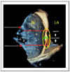
Segmental assessment of the mitral valve. (A) Mid-esophageal commissural view with biplane imaging allows systematic segmentation from the lateral commissure (noted by the left atrial appendage). (BD) Biplane cuts demonstrate sequential leaflet scallops: A1–P1 (red line, (B)), A2–P2 (yellow line, (C)), and A3–P3 (green line, (D)). (E) Three-dimensional “en face” view of the mitral valve from the left atrial perspective provides a comprehensive anatomical context and spatial orientation of scallops. LA: left atrium, LV: left ventricle, LAA: left atrial appendage, AoV: aortic valve.
Figure 5
Figure 5
(A) Posterior leaflet length measurement using 2D and (B) multiplanar reconstruction (MPR) at grasping zone. LA: left atrium, LV: left ventricle, AoV: aortic valve.
Figure 6
Figure 6
(A) Mitral valve area measurement using MPR. (B) Baseline mean mitral valve gradient using CW Doppler. MVA: mitral valve area.
Figure 7
Figure 7
(A,B) Measurement of flail width in primary MR (red line). (C,D) Measurement of flail gap (red line). MV: mitral valve, AoV: aortic valve.
Figure 8
Figure 8
(A) Measurement of coaptation depth (or tenting height) in secondary MR (red line). (B) Measurement of coaptation length (red line). (C) Measurement of coaptation gap (red line). MV mitral valve, AoV: aortic valve.
Figure 9
Figure 9
Three-dimensional “en face” mitral valve shows cleft-like indentation at the junction of the P2 and the P3 segment. (A) Atrial view or surgeon’s view and (B) ventricular view. LAA: left atrial appendage, AoV: aortic valve.
Figure 10
Figure 10
(A) Comparative assessment of posterior leaflet motion in (A) systolic and (B) diastolic phases. The posterior leaflet, indicated by a red arrow in both images, demonstrates a complete lack of movement throughout the cardiac cycle. LA: left atrium, LV: left ventricle, AML: anterior mitral leaflet, PML: posterior mitral leaflet.
Figure 11
Figure 11
(A) The posterior annulus (green arrow) is calcified and extended into the posterior leaflet, resulting in a shorter effective posterior leaflet for grasping in 2D image and (B) in 3D with multiplanar reconstruction. LA: left atrium, LV: left ventricle, AML: anterior mitral leaflet, PML: posterior mitral leaflet, LAA: left atrial appendage, AoV: aortic valve.
Figure 12
Figure 12
(A) The circle represents optimal transseptal puncture site (superior and posterior). (B) The short axis view guides anterior (towards the aorta) or posterior position, (C) the bi-caval view guides superior or inferior position, (D) the 4-chamber view is used for measurement of puncture height from the level of annulus and, (E) when the puncture site is positioned more superiorly than typically expected and the four-chamber view fails to adequately delineate both the puncture site and the annulus, a modified 135–150° view is employed to optimize alignment, thereby enabling precise visualization and measurement. LA: left atrium, LV: left ventricle, RA: right atrium, RV: right ventricle, AoV: aortic valve.
Figure 13
Figure 13
(A) Four-chamber view and (B) modified 135–150° view for measurement of the height of transeptal puncture from the level of annulus (vertical red line). LA: left atrium, LV: left ventricle, RA: right atrium, RV: right ventricle.
Figure 14
Figure 14
Superior puncture position offers better alignment to the lateral commissure, whereas inferior puncture position offers better alignment to the medial commissure. These adjustments are crucial to tailoring the procedure based on the targeted segment and ensuring optimal outcomes. LA: left atrium, LV: left ventricle, IAS: interatrial septum.
Figure 15
Figure 15
(A,B) Navigating the guiding catheter and the device towards the mitral valve. (C) Medial–lateral orientation (blue = lateral towards the left atrial appendage and green = medial towards the septum). (D) Anterior–posterior orientation (red = anterior towards the aortic valve and yellow = posterior). AoV: aortic valve, LAA: left atrial appendage, AML: anterior mitral leaflet, PML: posterior mitral leaflet.
Figure 16
Figure 16
Multi-planar reconstruction (MPR) for device orientation and perpendicularity. (A) Three-dimensional en-face view of the mitral valve, demonstrating device orientation, (B) short axis projection of the orientation of the device, (C) Bi-commissural view for medial-lateral position as-sessment of the device, (D) Long-axis view for anterior-posterior position assessment of the device. AoV: aortic valve, LAA: left atrial appendage, AML: anterior mitral leaflet, PML: posterior mitral leaflet.
Figure 17
Figure 17
Testing the grippers (indicated by red arrow) to ensure proper function. LA: left atrium, LV: left ventricle, AoV: aortic valve, AML: anterior mitral leaflet, PML: posterior mitral leaflet.
Figure 18
Figure 18
Three-dimensional mitral valve shows device orientation. (A) Perpendicularity control after crossing mitral valve to check optimal orientation. (B) After the device is advanced into the left ventricle (LV), its orientation can be assessed using 3D echocardiography. Reducing the gain during imaging enhances the clarity of the clip and its position, allowing for more precise visualization of its alignment in relation to the coaptation line. AoV: aortic valve, LAA: left atrial appendage.
Figure 19
Figure 19
(A) Failure to grasp anterior leaflet (indicated by red arrow), (B) partial capture of anterior leaflet (red arrow), (C) complete capture for both leaflets. AML: anterior mitral leaflet, PML: posterior mitral leaflet.
Figure 20
Figure 20
Post-device deployment echocardiographic assessment. (A) Color Doppler imaging reveals trace residual mitral regurgitation, indicating minimal leakage after device deployment. (B) Continuous-wave Doppler evaluation demonstrates a mean trans-mitral gradient of <5 mmHg, indicating the absence of significant iatrogenic mitral stenosis. (C) Three-dimensional multi-planar reconstruction confirms an adequate mitral valve area (MVA ≥ 1.5 cm2). (D) Three-dimensional image after device implantation demonstrates good tissue bridging. AoV: aortic valve, LAA: left atrial appendage, AML: anterior mitral leaflet, PML: posterior mitral leaflet, MVA: mitral valve area.
Figure 21
Figure 21
Residual leaflet prolapse after the deployment of the first device and subsequent correction with an additional device implanted medially to the first. (A) A 2D echocardiographic view demonstrates persistent leaflet prolapse, medially to the initial device. (B) The corresponding color Doppler image reveals a regurgitant jet at the medial aspect, indicating residual mild MR. (C) A 2D echocardiographic view, similar to (A), shows no residual prolapse after the placement of a second device, confirming optimal anatomical correction. (D) The corresponding color Doppler image, similar to (B), demonstrates non-significant residual MR. AML: anterior mitral leaflet, PML: posterior mitral leaflet, AoV: aortic valve.

References

    1. Nkomo V.T., Gardin J.M., Skelton T.N., Gottdiener J.S., Scott C.G., Enriquez-Sarano M. Burden of valvular heart diseases: A population-based study. Lancet. 2006;368:1005–1011. doi: 10.1016/S0140-6736(06)69208-8. - DOI - PubMed
    1. Iung B., Baron G., Butchart E.G., Delahaye F., Gohlke-Bärwolf C., Levang O.W., Tornos P., Vanoverschelde J.-L., Vermeer F., Boersma E., et al. A prospective survey of patients with valvular heart disease in Europe: The Euro Heart Survey on Valvular Heart Disease. Eur. Heart J. 2003;24:1231–1243. doi: 10.1016/S0195-668X(03)00201-X. - DOI - PubMed
    1. Zoghbi William A., Levine Robert A., Flachskampf F., Grayburn P., Gillam L., Leipsic J., Thomas James D., Kwong Raymond Y., Vandervoort P., Chandrashekhar Y. Atrial Functional Mitral Regurgitation. JACC Cardiovasc. Imaging. 2022;15:1870–1882. doi: 10.1016/j.jcmg.2022.08.016. - DOI - PubMed
    1. Iliakis P., Dimitriadis K., Pyrpyris N., Beneki E., Theofilis P., Tsioufis P., Kamperidis V., Aznaouridis K., Aggeli K., Tsioufis K. Atrial Functional Mitral Regurgitation: From Diagnosis to Current Interventional Therapies. J. Clin. Med. 2024;13:5035. doi: 10.3390/jcm13175035. - DOI - PMC - PubMed
    1. Prakash R., Horsfall M., Markwick A., Pumar M., Lee L., Sinhal A., Joseph M.X., Chew D.P. Prognostic impact of moderate or severe mitral regurgitation (MR) irrespective of concomitant comorbidities: A retrospective matched cohort study. BMJ Open. 2014;4:e004984. doi: 10.1136/bmjopen-2014-004984. - DOI - PMC - PubMed

LinkOut - more resources