

Cardiac Magnetic Resonance Imaging and Arrhythmic Risk Stratification in Cardiomyopathies
- PMID: 40725614
- PMCID: PMC12295573
- DOI: 10.3390/jcm14144922
Cardiac Magnetic Resonance Imaging and Arrhythmic Risk Stratification in Cardiomyopathies
Abstract
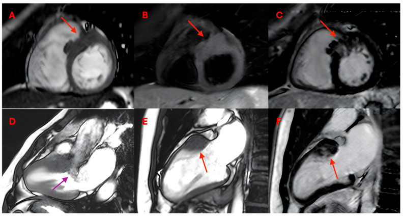
Cardiac magnetic resonance imaging (CMRI) has become an indispensable tool in evaluating arrhythmic risk and guiding therapeutic decisions in patients with non-ischemic cardiomyopathies (NICMs), including dilated (DCM), hypertrophic (HCM), and arrhythmogenic cardiomyopathies (ACM). Both European and American guidelines have given an additive and different value of late gadolinium enhancement (LGE) in specific morpho-functional (hypertrophic, dilated, and arrhythmogenic) phenotypes. In particular, LGE plays a different weight in relation to different cardiomyopathies. In dilated cardiomyopathy, LGE is able to predict arrhythmic risk in relationship to the presence and localization (septal and/or ring like LGE). On the contrary, in HCM, LGE is related to increased risk of cardiac death according to the extent (LGE >15%), while in ACM, it has a greater role in the presence of fat infiltration associated with LGE. In this review, we aim to identify predictors of sudden cardiac death related to myocardial structural features seen in CMRI in cardiomyopathies, going beyond the sole assessment of left ventricular function and ejection fraction.
Keywords: cardiac imaging; cardiac magnetic resonance imaging; cardiomyopathies; sudden cardiac death; ventricular arrhythmias.
Conflict of interest statement
The authors declare no conflicts of interest.
Figures


Similar articles
-
Late Gadolinium Enhancement and the Risk for Ventricular Arrhythmias or Sudden Death in Dilated Cardiomyopathy: Systematic Review and Meta-Analysis.JACC Heart Fail. 2017 Jan;5(1):28-38. doi: 10.1016/j.jchf.2016.09.017. Epub 2016 Dec 21. JACC Heart Fail. 2017. PMID: 28017348
-
Magnetic Resonance Imaging Characterization and Clinical Outcomes of Dilated and Arrhythmogenic Left Ventricular Cardiomyopathies.J Am Coll Cardiol. 2024 May 14;83(19):1841-1851. doi: 10.1016/j.jacc.2024.02.041. J Am Coll Cardiol. 2024. PMID: 38719365 Free PMC article.
-
Prognostic impact of late gadolinium enhancement granularity in non-ischemic dilated cardiomyopathy.Eur Radiol. 2025 Aug;35(8):4699-4710. doi: 10.1007/s00330-025-11404-8. Epub 2025 Feb 8. Eur Radiol. 2025. PMID: 39920302
-
Prognostic value of late gadolinium enhancement in clinical outcomes for hypertrophic cardiomyopathy.JACC Cardiovasc Imaging. 2012 Apr;5(4):370-7. doi: 10.1016/j.jcmg.2011.11.021. JACC Cardiovasc Imaging. 2012. PMID: 22498326
-
Late gadolinium enhancement dispersion for predicting malignant arrhythmic events in patient with non-ischaemic dilated cardiomyopathy.Eur Heart J Cardiovasc Imaging. 2025 Jun 30;26(7):1217-1232. doi: 10.1093/ehjci/jeaf124. Eur Heart J Cardiovasc Imaging. 2025. PMID: 40244913
References
-
- Hammersley D.J., Zegard A., Androulakis E., Jones R.E., Okafor O., Hatipoglu S., Mach L., Lota A.S., Khalique Z., de Marvao A., et al. Arrhythmic Risk Stratification by Cardiovascular Magnetic Resonance Imaging in Patients with Nonischemic Cardiomyopathy. J. Am. Coll. Cardiol. 2024;84:1407–1420. doi: 10.1016/j.jacc.2024.06.046. - DOI - PMC - PubMed
-
- Licordari R., Trimarchi G., Teresi L., Restelli D., Lofrumento F., Perna A., Campisi M., de Gregorio C., Grimaldi P., Calabrò D., et al. Cardiac Magnetic Resonance in HCM Phenocopies: From Diagnosis to Risk Stratification and Therapeutic Management. J. Clin. Med. 2023;12:3481. doi: 10.3390/jcm12103481. - DOI - PMC - PubMed
-
- Theerasuwipakorn N., Chokesuwattanaskul R., Phannajit J., Marsukjai A., Thapanasuta M., Klem I., Chattranukulchai P. Impact of late gadolinium-enhanced cardiac MRI on arrhythmic and mortality outcomes in nonischemic dilated cardiomyopathy: Updated systematic review and meta-analysis. Sci. Rep. 2023;13:13775. doi: 10.1038/s41598-023-41087-4. - DOI - PMC - PubMed
-
- Eichhorn C., Koeckerling D., Reddy R.K., Ardissino M., Rogowski M., Coles B., Hunziker L., Greulich S., Shiri I., Frey N., et al. Risk Stratification in Nonischemic Dilated Cardiomyopathy Using CMR Imaging: A Systematic Review and Meta-Analysis. JAMA. 2024;332:1535–1550. doi: 10.1001/jama.2024.13946. - DOI - PMC - PubMed
Publication types
LinkOut - more resources
Full Text Sources
