

Assessment of Retinal Microcirculation in Primary Open-Angle Glaucoma Using Adaptive Optics and OCT Angiography: Correlation with Structural and Functional Damage
- PMID: 40725669
- PMCID: PMC12296177
- DOI: 10.3390/jcm14144978
Assessment of Retinal Microcirculation in Primary Open-Angle Glaucoma Using Adaptive Optics and OCT Angiography: Correlation with Structural and Functional Damage
Abstract
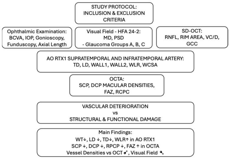
Background: This study aimed to evaluate retinal arteriole parameters using adaptive optics (AO) rtx1™ (Imagine Eyes, Orsay, France) and peripapillary and macular vessel densities with optical coherence tomography angiography (OCTA) in eyes with different stages of primary open-angle glaucoma (POAG) compared to healthy eyes. It also investigated the associations between vascular parameters and glaucoma severity, as defined by structural (OCT) and functional (visual field) changes. Methods: Fifty-seven eyes from 31 POAG patients and fifty from 25 healthy volunteers were examined. Retinal arteriole morphology was assessed using the AO rtx1™-fundus camera, which measured lumen diameter, wall thickness, total diameter, wall-to-lumen ratio (WLR), and wall cross-sectional area. OCTA was used to measure vessel densities in superficial (SCP) and deep (DCP) capillary plexuses of the macula and radial peripapillary capillary plexus (RPCP) and FAZ area. Structural OCT parameters (RNFL, GCC, rim area) and visual field tests (MD, PSD) were also performed. Results: Glaucoma eyes showed significantly thicker arteriole walls (12.8 ± 1.4 vs. 12.2 ± 1.3 µm; p = 0.030), narrower lumens (85.5 ± 10.4 vs. 100.6 ± 11.1 µm; p < 0.001), smaller total diameters (111.0 ± 10.4 vs. 124.1 ± 12.4 µm; p < 0.001), and higher WLRs (0.301 ± 0.04 vs. 0.238 ± 0.002; p < 0.001) than healthy eyes. In glaucoma patients, OCTA revealed significantly reduced vessel densities in SCP (36.39 ± 3.60 vs. 38.46 ± 1.41; p < 0.001), DCP (36.39 ± 3.60 vs. 38.46 ± 1.41; p < 0.001), and RPCP plexuses (35.42 ± 4.97 vs. 39.27 ± 1.48; p < 0.001). The FAZ area was enlarged in eyes with glaucoma (0.546 ± 0.299 vs. 0.295 ± 0.125 mm2); p < 0.001). Positive correlations were found between vessel densities and OCT parameters (RNFL, r = 0.621; GCC, r = 0.536; rim area, r = 0.489), while negative correlations were observed with visual field deficits (r = -0.517). Conclusions: Vascular deterioration, assessed by AO rtx1™ and OCTA, correlates closely with structural and functional damage in glaucoma. Retinal microcirculation changes may precede structural abnormalities in the optic nerve head. Both imaging methods enable the earlier detection, staging, and monitoring of glaucoma compared to conventional tests.
Keywords: adaptive optics; foveal avascular zone; ganglion cell complex; optical coherence tomography angiography; primary open-angle glaucoma; retinal arteriole morphology; retinal microcirculation; retinal nerve fibre layer; vessel density.
Conflict of interest statement
The authors declare no conflicts of interest.
Figures
Similar articles
-
Diagnostic Capability of OCTA-Derived Macular Biomarkers for Early to Moderate Primary Open Angle Glaucoma.J Clin Med. 2024 Jul 18;13(14):4190. doi: 10.3390/jcm13144190. J Clin Med. 2024. PMID: 39064230 Free PMC article.
-
Optical coherence tomography and angiography reveal early retinal alterations in pediatric-onset multiple sclerosis.Eur J Pediatr. 2025 Jun 6;184(7):398. doi: 10.1007/s00431-025-06234-1. Eur J Pediatr. 2025. PMID: 40478274
-
Assessment of Vascular Density, Macular Layer Thickness, and Foveal Avascular Zone in Pseudoexfoliation Disease.J Glaucoma. 2025 Aug 19. doi: 10.1097/IJG.0000000000002620. Online ahead of print. J Glaucoma. 2025. PMID: 40833342
-
Optic nerve head and fibre layer imaging for diagnosing glaucoma.Cochrane Database Syst Rev. 2015 Nov 30;2015(11):CD008803. doi: 10.1002/14651858.CD008803.pub2. Cochrane Database Syst Rev. 2015. PMID: 26618332 Free PMC article.
-
Comprehensive assessment of glaucoma in patients with high myopia: a systematic review and meta-analysis with a discussion of structural and functional imaging modalities.Int Ophthalmol. 2024 Oct 11;44(1):405. doi: 10.1007/s10792-024-03321-4. Int Ophthalmol. 2024. PMID: 39392516 Free PMC article.
References
LinkOut - more resources
Full Text Sources
Miscellaneous
