

18F-FDG PET/CT identifies isolated metastasis of renal cancer in a patient with end-stage renal disease: A case report
- PMID: 40725905
- PMCID: PMC12303487
- DOI: 10.1097/MD.0000000000043595
18F-FDG PET/CT identifies isolated metastasis of renal cancer in a patient with end-stage renal disease: A case report
Abstract
Rationale: Individuals with end-stage renal disease have a considerably higher rate of malignant tumors, especially renal cancer, in comparison to the general population. However, cases in which bone metastases in the humerus are the initial clinical presentation that results in a diagnosis of renal cancer are extremely rare. Diagnosing renal cancer in patients with end-stage renal disease can be challenging when the clinical symptoms are atypical and do not present the "renal cancer triad." Our case report highlights the diagnostic importance of positron emission tomography/computed tomography (CT) imaging, increases clinicians' awareness of the disease, explores the potential etiology of renal cancer associated with end-stage renal disease, and provides insights into diagnostic and therapeutic strategies.
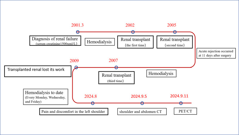
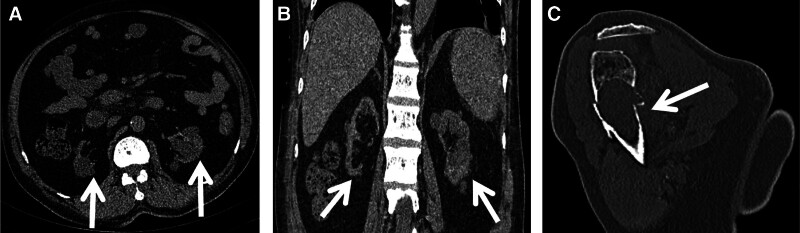
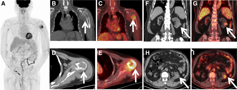
Patient concerns: A 58-year-old man, who has undergone multiple renal transplants and maintenance dialysis therapy for over 20 years due to renal failure, arrived at our hospital with complaints of left shoulder pain and progressive worsening. CT revealed unexplained osteolytic destruction and fracture in the left humerus. The 18F-fluorodeoxyglucose positron emission tomography/CT scan detected soft tissue lesions in the left kidney and exhibited increased fluorodeoxyglucose uptake.
Diagnoses: Upon conducting a biopsy on the patient's left humerus, metastatic renal cancer was diagnosed.
Interventions: The patient selected conservative treatment, and the injured humerus was immobilized to stabilize the affected area. There was no additional active treatment for renal cancer.
Outcomes: As of the time of submission, the patient's pain had markedly intensified, requiring oral pain medication for symptomatic relief.
Lessons: It is indeed rare for bone metastasis in the humerus to be the initial clinical sign that leads to the diagnosis of renal cancer in patients with end-stage renal disease. Through this case report, we aimed to enhance awareness and deepen understanding of renal cancer associated with end-stage renal disease.
Keywords: PET/CT; end-stage renal disease; renal cancer.
Copyright © 2025 the Author(s). Published by Wolters Kluwer Health, Inc.
Conflict of interest statement
The authors have no funding and conflicts of interest to disclose.
Figures
Similar articles
-
123I-MIBG scintigraphy and 18F-FDG-PET imaging for diagnosing neuroblastoma.Cochrane Database Syst Rev. 2015 Sep 29;2015(9):CD009263. doi: 10.1002/14651858.CD009263.pub2. Cochrane Database Syst Rev. 2015. PMID: 26417712 Free PMC article.
-
Fluorine-18-fluorodeoxyglucose (FDG) positron emission tomography (PET) computed tomography (CT) for the detection of bone, lung, and lymph node metastases in rhabdomyosarcoma.Cochrane Database Syst Rev. 2021 Nov 9;11(11):CD012325. doi: 10.1002/14651858.CD012325.pub2. Cochrane Database Syst Rev. 2021. PMID: 34753195 Free PMC article.
-
¹⁸F-FDG PET/CT: a review of diagnostic and prognostic features in multiple myeloma and related disorders.Clin Exp Med. 2015 Feb;15(1):1-18. doi: 10.1007/s10238-014-0308-3. Epub 2014 Sep 14. Clin Exp Med. 2015. PMID: 25218739
-
Positron emission tomography-adapted therapy for first-line treatment in individuals with Hodgkin lymphoma.Cochrane Database Syst Rev. 2015 Jan 9;1(1):CD010533. doi: 10.1002/14651858.CD010533.pub2. Cochrane Database Syst Rev. 2015. Update in: Cochrane Database Syst Rev. 2025 Mar 26;3:CD010533. doi: 10.1002/14651858.CD010533.pub3. PMID: 25572491 Free PMC article. Updated.
-
The value of FDG positron emission tomography/computerised tomography (PET/CT) in pre-operative staging of colorectal cancer: a systematic review and economic evaluation.Health Technol Assess. 2011 Sep;15(35):1-192, iii-iv. doi: 10.3310/hta15350. Health Technol Assess. 2011. PMID: 21958472 Free PMC article.
References
-
- Neuzillet Y, Tillou X, Mathieu R, et al. ; Comité de Transplantation de l'Association Française d'Urologie. Renal cell carcinoma (RCC) in patients with end-stage renal disease exhibits many favourable clinical, pathologic, and outcome features compared with RCC in the general population. Eur Urol. 2011;60:366–73. - PubMed
Publication types
MeSH terms
Substances
LinkOut - more resources
Full Text Sources
Medical
Research Materials
