Review
. 2025 Jul 28:19417381251352500.
doi: 10.1177/19417381251352500. Online ahead of print.

Magnetic Resonance Neurography of Sports-Related Peripheral Nerve Injuries of the Shoulder Region

Affiliations
Review

Magnetic Resonance Neurography of Sports-Related Peripheral Nerve Injuries of the Shoulder Region

Yenpo Lin et al. Sports Health. .

Abstract

Context: Although many sports-related injuries in the shoulder region are self-limiting, those involving the peripheral nerves can have a profound adverse effect on an athlete's career. While physical examination and electrodiagnostic testing are the mainstays for assessing such injuries, peripheral nerve magnetic resonance (MR) imaging, also known as MRN, can play an important and complementary role in helping further characterize injury extent and localization. This review explores the application of MRN in diagnosing and managing sports-related peripheral nerve injuries.

Evidence acquisition: A narrative overview of the literature combined with the clinical and research expertise of the authors.

Study design: Narrative review.

Level of evidence: Level 4.

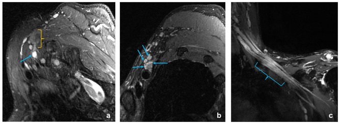
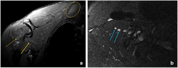
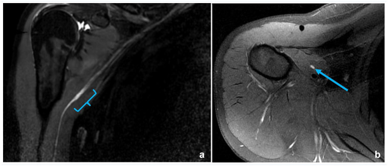
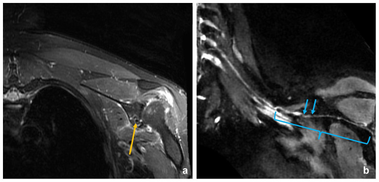
Results: MRN helps localize preganglionic and postganglionic traumatic brachial plexus injuries, facilitating the planning of appropriate surgical interventions and determining the extent of nerve injury. MRN may also be used to assess thoracic outlet syndrome, commonly implicated in overhead sports activities. In cases of shoulder dislocation, MRN can depict altered nerve signal intensity and morphology of the axillary nerve that may be stretched. MRN also plays an important role in detecting and guiding the management of iatrogenic nerve-related injuries, and helping define alternative diagnoses, such as Parsonage-Turner syndrome, that might be the culprit in the postsurgical setting. Bone-sensitive and angiography MR pulse sequences can help delineate the spatial relationship among peripheral nerves, osseous structures, and blood vessels.

Conclusion: MRN is valuable in assessing sports-related traumatic nerve injuries of the shoulder region, as well as newly developed neurological symptoms following shoulder surgery. Specifically, MRN provides a global evaluation of regional muscles and nerves, which and can help localize injury to one or more nerve segments that may be targeted for therapeutic intervention.Strength-of-Recommendation Taxonomy (SORT):Evidence B level 3.

Keywords: MR neurography; brachial plexus injury; peripheral nerve injury; thoracic outlet syndrome.


Conflict of interest statement

The following authors declared potential conflicts of interest: E.T.T. has received institutional research support from GE Healthcare, Siemens Healthcare, Medtronic, and Azurity Pharmaceuticals and the HSS Innovation Institute; provisional patents in system and apparatus for overlapping phased array coils for curved surfaces, system and method for MR neurography, and system and apparatus for simplified diffusion imaging. D.B.S. has institutional research agreements with GE Healthcare and Siemens Medical Solutions; and has received research support from Azurity Pharmaceuticals and the HSS Innovation Institute; consultant, GE Healthcare; provisional patents in system and apparatus for overlapping phased-array coils for curved surfaces, system and method for MR neurography, and system and apparatus for simplified diffusion imaging.

The authors disclosed receipt of the following financial support for the research, authorship, and/or publication of this article: National Institute for Biomedical Imaging and Bioengineering, NIH R01EB031820.

Figures

Figure 1.
Figure 1.
Magnetic resonance neurography of the brachial plexus. (a) Maximal intensity projection coronal short-tau inversion recovery image demonstrates normal appearance of the BP. (b) Oblique sagittal T2-weighted fat-suppressed image, acquired at the level of the dashed line in a, shows normal fascicular architecture of the lateral (blue arrow), posterior (green arrow), and medial (yellow arrow) cords.
Figure 2.
Figure 2.
A 46-year-old man with persistent neck and shoulder pain for 7 months after a jujitsu injury. (a,b) Oblique sagittal T2-weighted fat-suppressed images reveal hyperintensity and enlargement of the C7 nerve root (arrow in a) and middle scalene muscle edema (bracket in a). Additional hyperintensity and enlargement are seen in the upper and middle trunks and divisions (arrows in b). (c) Coronal short-tau inversion recovery image further confirms BP involvement (bracket), suggestive of stretch injury. BP, brachial plexus.
Figure 3.
Figure 3.
A 22-year-old man with severe weakness and numbness of the left upper arm after a motorcycle racing injury 6 months previously. (a) Coronal T2-weighted fat-suppressed image shows denervation of the deltoid (*), supraspinatus (^), subscapularis (@), and cervical paraspinal (ellipse) muscles. (b,c) Oblique sagittal T2-weighted fat-suppressed images reveal a C8 pseudomeningocele (arrow in b) as well as enlarged UT, MT, and C8 and T1 nerve roots (arrows in c). (d) Coronal short-tau inversion recovery image demonstrates failure to identify the extraforaminal C5 and C6 nerve roots (bracket), compatible with rupture, along with hyperintense and enlarged divisions and cords (arrows). UT, upper trunk; MT, middle trunk.
Figure 4.
Figure 4.
A 25-year-old male American football cornerback with right arm weakness and paresthesias after a ‘hit’ 2 days previously. (a) Coronal T2-weighted fat-suppressed image shows mild edema patterns in the right serratus anterior (arrow), teres minor (dashed arrow), and right paraspinal musculature (ellipse), which could reflect muscle strain. (b) Oblique sagittal T2-weighted fat-suppressed image shows very mild signal hyperintensity of C5 and C6 nerve roots (arrows), potentially indicating a stretch injury.
Figure 5.
Figure 5.
A 30-year-old female yogi with paresthesias in both hands, right greater than left. (a) The frontal cervical spine radiograph shows a prominent, right cervical rib (bracket). (b) Oblique sagittal fat-suppressed image shows enlargement and hyperintensity of the right BP divisions (ellipse). (c,d) MRA image with both the arms elevated (c) and in the neutral position (d) reveals severe narrowing of the proximal to midsegment of the right subclavian artery (bracket in c) during arm elevation, likely related to compression by the cervical rib. BP, brachial plexus; MRA, magnetic resonance angiography.
Figure 6.
Figure 6.
A 21-year-old male professional rugby player presenting with right upper arm pain and weakness. (a) Coronal T2 fat-suppressed image demonstrates diffuse denervation pattern of the deltoid muscle (*) and teres minor (^). (b) Coronal short-tau inversion recovery image reveals prominent signal hyperintensity of the right axillary nerve as it leaves the posterior cord (bracket), suggestive of stretch injury.
Figure 7.
Figure 7.
A 27-year-old man with left shoulder dislocation after sandboarding injury 10 months previously. (a) Coronal T2-weighted fat-suppressed image shows diffuse edema pattern and reduced bulk of the deltoid (*) and teres minor (^) muscles. (b) Coronal short-tau inversion recovery image demonstrates a ruptured axillary nerve immediately after it leaves the posterior cord with a recoiled terminal neuroma (arrows). (c) A fused-rendering image from (b) and a ZTE sequence demonstrate the spatial relationship of the neuroma (arrow) to the coracoid process (dashed arrow). ZTE, zero-echo-time.
Figure 8.
Figure 8.
A 35-year-old man with left shoulder pain and weakness after mountain-climbing with a heavy backpack. (a) Coronal T2-weighted fat-suppressed image shows denervation edema of the infraspinatus (*) but with sparing of the supraspinatus (^). (b) Axial intermediate-weighted image shows a ganglion cyst (arrow) at the spinoglenoid notch. (c) Maximal intensity projection coronal short-tau inversion recovery image demonstrates hyperintensity and enlargement of the distal segment (bracket) of the suprascapular nerve, impinged upon by the ganglion cyst (arrow).
Figure 9.
Figure 9.
A 27-year-old male professional baseball pitcher presenting with numbness and paresthesias in the right ring finger for 1 week. (a) Axial intermediate-weighted image demonstrates ill-defined high signal surrounding the axillary nerve and circumflex vessels (bracket). (b) Maximum intensity projection MRA image reveals abrupt narrowing of the origin of the posterior humeral circumflex artery (arrow), suggestive of intimal injury with subsequent distal thrombosis. MRA, magnetic resonance angiography.
Figure 10.
Figure 10.
A 23-year-old female softball pitcher presented with popping sensation and subsequent biceps weakness and lateral forearm numbness. (a) Coronal short-tau inversion recovery image demonstrates short segment signal hyperintensity and enlargement of the musculocutaneous nerve (bracket) in the proximal aspect of the short head biceps/coracobrachialis muscles. (b) Oblique axial T2-weighted fat-suppressed image redemonstrates the hyperintensity and enlargement of the musculocutaneous nerve (arrow).
Figure 11.
Figure 11.
A 43-year-old male presented with left arm weakness immediately after left shoulder capsulolabral repair. (a) Coronal T2-weighted fat-suppressed image shows denervation edema of the deltoid (*) and teres minor (^) muscles. (b) Oblique sagittal intermediate-weighted image demonstrates enlargement of the axillary nerve (bracket) with perineural scarring in close proximity to the surgical materials (arrows). (c) Coronal short-tau inversion recovery image demonstrates hyperintensity and enlargement of the axillary nerve as it leaves the posterior cord (blue bracket), suggesting neuroma-in-continuity. Distal to this, a nerve segment (red bracket) is poorly visualized, likely reflecting scar encasement. The distal segment (yellow bracket) appears normal.
Figure 12.
Figure 12.
A 17-year-old female soccer goalie with grade 3 AC joint sprain presented with severe weakness in external rotation 4 months after arthroscopic coracoclavicular ligament allograft reconstruction. (a) Radiograph of the right AC joint shows the implant (arrow) in the midclavicle. (b) Coronal T2-weighted fat-suppressed image shows denervation edema and reduced bulk of the supraspinatus (*) and infraspinatus (^) muscles. (c) Coronal T2-weighted fat-suppressed image shows enlargement and hyperintensity of the suprascapular nerve (blue arrow) proximal to the graft (yellow arrow). (d) Axial T2-weighted fat-suppressed image demonstrates anterior deviation of the suprascapular nerve (blue arrow), which is tethered by the graft (yellow arrow). AC, acromioclavicular.
Figure 13.
Figure 13.
A 35-year-old man presented with shoulder pain and paresthesias 5 days after arthroscopic rotator cuff and Bankart lesion repair for a snowboarding injury. (a) Coronal T2-weighted fat-suppressed image demonstrates a suture anchor (yellow arrow) in the glenoid. (b) Curved multiplanar reconstructed image of coronal short-tau inversion recovery fast spin echo sequence reveals 2 sites of focal caliber change (arrows) of the suprascapular nerve (bracket), which is often seen in PTS. PTS, Parsonage-Turner syndrome.

Similar articles

References

    1. AlMakadma Y, Eirale C, Chamari K. Neuropathic pain in athletes: basics of diagnosis and monitoring of a hidden threat. Biol Sport. 2022;39(4):943-949. PMID: 36247942 - PMC - PubMed
    1. Aragaki D, Basu A, Conlon C, et al. Quality of electrodiagnostic testing for carpal tunnel syndrome: adherence to quality measures. Muscle Nerve. 2020;62(1):50-59. PMID: 32133653 - PubMed
    1. Avis D, Power D. Axillary nerve injury associated with glenohumeral dislocation. EFORT Open Rev. 2018;3(3):70-77. PMID: 29657847 - PMC - PubMed
    1. Bowles DR, Canseco JA, Alexander TD, Schroeder GD, Hecht AC, Vaccaro AR. The prevalence and management of stingers in college and professional collision athletes. Curr Rev Musculoskelet Med. 2020;13(6):651-662. PMID: 32691363 - PMC - PubMed
    1. Carvalho GA, Nikkhah G, Matthies C, Penkert G, Samii M. Diagnosis of root avulsions in traumatic brachial plexus injuries: value of computerized tomography myelography and magnetic resonance imaging. J Neurosurg. 1997;86(1):69-76. PMID: 8988084 - PubMed

LinkOut - more resources