. 2025 Nov;80(11):3095-3107.
doi: 10.1111/all.16682. Epub 2025 Jul 29.

Distinct Roles for Thymic Stromal Lymphopoietin (TSLP) and IL-33 in Experimental Eosinophilic Esophagitis

Affiliations

Distinct Roles for Thymic Stromal Lymphopoietin (TSLP) and IL-33 in Experimental Eosinophilic Esophagitis

Anish Dsilva et al. Allergy. 2025 Nov.

Abstract

Rationale: Thymic stromal lymphopoietin (TSLP) and IL-33 are alarmins implicated in eosinophilic esophagitis (EoE) pathogenesis by activating multiple cells, including mast cells (MCs). Whether TSLP or IL-33 have a role in EoE and whether their activities are distinct requires further investigation.

Methods: Experimental EoE was induced in wild type (WT) Il33-/- and Crlf2-/- mice. TSLP or IL-5 were neutralized using antibodies. Esophageal histopathology was determined by H&E, anti-Ki67, anti-CD31, and anti-MBP staining. Esophageal RNA was subjected to RNA sequencing. Bone marrow-derived MCs were activated with TSLP and IL-13 was determined (ELISA).

Results: TSLP and IL-33 were overexpressed in human and experimental EoE. Human and mouse esophageal MCs displayed the highest level of Crlf2 (TSLPR) compared to other immune cells. Crlf2-/- mice were nearly completely protected from EoE, and TSLP neutralization resulted in decreased basal cell proliferation, eosinophilia, lamina propria thickening, and vascularization. Induction of experimental EoE in Il33-/- mice resulted in reduced eosinophilia, but no alterations in tissue remodeling were observed compared to WT mice. RNA sequencing revealed that TSLP regulates the expression of key genes associated with human EoE (e.g., eotaxins, Il19, Klk5, Flg, Il36rn, Il1r2) and suggests a role for TSLP in regulating IL-1 signaling, barrier integrity, and epithelial cell differentiation. Experimental EoE was characterized by a MC-associated gene signature and elevated MCs. Activation of MCs with TSLP resulted in the secretion of IL-13.

Conclusion: TSLP and IL-33 have non-redundant functions in experimental EoE. This study highlights TSLP as an upstream regulator of IL-13 and a potential therapeutic target for EoE.

Keywords: IL‐33; TSLP; allergy; eosinophilic esophagitis.


Figures

Figure 1.
Figure 1.. Expression of TSLP, IL-33 and their receptors in experimental and human EoE
Schematic representation of the experimental EoE model (A). Kinetic analysis of TSLP (black circle) and IL-33 (white circle) expression in the esophagus (B) and ear (C) throughout the experimental EoE protocol. Esophageal samples were obtained and the expression of IL-33 and TSLP were determined at day 36 by immunohistochemistry or immunofluorescence, respectively (D, F). Thereafter, the number of IL-33+ cells or the density of TSLP staining in the esophagus were quantified (E, G). Analysis of IL-33 and TSLP expression in human EoE samples was obtained from the human single cell portal (H, I). The expression of mouse IL1RL1 (ST2) and CRLF2 was determined in single cell suspensions obtained from the esophagus (J, K) using polychromatic flow cytometry. Data are shown as fold increase of anti-ST2 (J) or anti-CRLF2 (K) over isotype control. The red line represents no fold induction when calculating anti-ST2 or anti-CRLF2 over isotype control. Analysis of Il1rl1 and Crlf2 expression in human EoE samples was obtained from the human single cell portal (L, M). Cells in the portal were annotated using the single cell portal (https://singlecell.broadinstitute.org/single_cell). In B, C, data are presented as mean ± SEM from one experiment with n= 6–15 mice per time point. In E, G, each dot represents one mouse from day 36. In H, I, L and M each dot represents one cell where the intensity of the color represents the expression of Il33 or Tslp. *- p<0.05, **- p<0.01, ***- p<0.001.
Figure 2.
Figure 2.. IL-33 regulates the accumulation of eosinophils in experimental EoE.
Experimental EoE was induced in wild type (WT) and Il33−/− mice using oxazolone (OXA). On day 36, the mice were euthanized, and esophageal tissues were fixed, paraffin embedded, and slides were generated. The slides were stained with anti-major basic protein (MBP, A) and esophageal eosinophils were quantified (B). Epithelial cell proliferation was determined using anti-Ki67 staining (C, D). H&E-stained slides were analysed for lamina propria (E-F) and esophageal epithelial (G) thickness. Esophageal vascularization was determined using anti-CD31 staining (H, I). Representative photomicrographs of anti-MBP (A), anti-Ki-67 (C), H&E (F) and anti-CD31 (H) are shown. Data are presented as mean ± SEM and are representative of one out of 3 different experiments conducted with 10–12 mice per group, ns-nonsignificant **-p<0.01, ***-p<0.001, ****- p<0.0001.
Figure 3.
Figure 3.. Experimental EoE is critically regulated by TSLP receptor
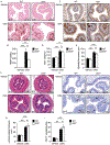
Experimental EoE was induced in wild type (WT) and Crlf2−/− mice using oxazolone (OXA). On day 36, the mice were euthanized, and esophageal tissues were fixed, paraffin embedded, and slides were generated. The slides were stained with anti-major basic protein (MBP, A) and esophageal eosinophils were quantified (B). Epithelial cell proliferation was determined using anti-Ki67 staining (C, D). H&E-stained slides were analysed for assessment of epithelial thickness (E-F) and lamina propria thickness (G). Esophageal vascularization was determined using anti-CD31 staining (H, I). Expression of IL-33+ cells was determined using anti-IL-33 immunohistochemistry (J). Experimental EoE was induced in wild type (WT) and Il33−/− mice using oxazolone (OXA). Expression of TSLP was determined using anti-TSLP by immunofluorescence (K-L). Representative photomicrographs of anti-MBP (A), anti-Ki-67 (C), H&E (F) and anti-CD31 (H) and anti-TSLP (K) are shown. Data are presented as mean ± SEM and are representative of one out of 3 different experiments conducted with 10–12 mice per group, ns-nonsignificant **-p<0.01, ***-p<0.001, ****- p<0.0001.
Figure 4.
Figure 4.. Pharmacological blockade of TSLP regulates eosinophilic infiltration, epithelial cell proliferation and esophageal vascularization in EoE.
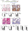
Experimental EoE was induced in wild type (WT) mice using oxazolone (OXA). Starting on day 19, the mice received two intraperitoneal injections per week of isotype control antibodies or anti-TSLP (clones M702 or clone 28F12, A). On day 36, the mice were euthanized, and esophageal tissues were fixed, paraffin embedded, and slides were generated. The slides were stained with anti-major basic protein (MBP, B) and esophageal eosinophils were quantified (C). Epithelial cell proliferation was determined using anti-Ki67 staining (D, E). H&E-stained slides (G) were analysed for lamina propria (F) as well as epithelial (H) thickness. Esophageal vascularization was determined using anti-CD31 staining (I, J). Single cell suspensions of esophageal tissue were obtained from oxazolone-challenged isotype control or anti-TSLP (M702)-treated mice using enzymatic digestion and the levels of eosinophils (K), CD4+ T cells (L) and mast cells (M) determined by flow cytometry. Representative photomicrographs of anti-MBP (B), anti-Ki-67 (D), H&E (G) and anti-CD31 (I) are shown. Data are presented as mean ± SEM and are representative of one out of 3 different experiments conducted with 10–12 mice per group, ns-nonsignificant, *- p<0.05, **-p<0.01, ***-p<0.001, ****- p<0.0001.
Figure 5.
Figure 5.. RNA sequencing of anti-TSLP-treated mice
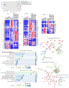
Experimental EoE was induced in wild type (WT) mice. Starting on day 19, the mice received two intraperitoneal injections per week of isotype control antibodies or anti-TSLP (clones M702 or clone 28F12). On day 36, the mice were euthanized, and esophageal tissue was obtained and subjected to bulk RNA sequencing. PCA plot of vehicle- and oxazolone (OXA)-challenged mice treated with either isotype control or anti-TSLP antibodies (clones M702 and/or 28F12) is shown (A). Heat plot analysis of all differentially expressed genes (absolute fold change > 2, p<0.05) (B) is presented. Analysis of the top differentially expressed secreted factors, cytokine receptors, adhesion molecules and extracellular matrix components is shown (C-D). Gene Set Enrichment Analysis (GSEA) and STRING analysis were performed on the DEGs, and comparison of OXA-challenged vehicle-treated mice vs. OXA-challenged anti-TSLP treated mice was performed (E-H). The top up and down regulated pathways, which were regulated by TSLP are presented (E-F). In C, D and E, each lane represents a different mouse.
Figure 6.
Figure 6.. Experimental EoE is characterized by a mucosal mast cell that secrete IL-13 upon stimulation with TSLP
WT mice were treated with vehicle or oxazolone (OXA) to induce experimental EoE. On day 36, the mice were euthanized, and esophageal tissue was obtained and subjected to bulk RNA sequencing. Analysis of the top ten upregulated proteases is shown (p < 0.02, >2-fold, A). Single-cell suspensions were prepared from enzymatically digested esophageal samples and stained with DAPI, anti-CD45, anti–c-Kit, and anti-FcεR1. Thereafter, the percentage of c-Kit+/FcεR1+ cells were determined (B). Bone marrow-derived mast cells (MCs) were grown and differentiated into a mucosal or connective tissue phenotype (C-D). Thereafter, the MCs were stimulated with TSLP (50 ng/mL) and the secretion of IL-13 was determined by ELISA (C-D). Data are from n = 3. Data are representative of n = 3 conducted in 3–5 technical repeats, ns- nonsignificant, *- p < 0.05.

Update of

References

    1. O’Shea KM, Aceves SS, Dellon ES, et al. Pathophysiology of Eosinophilic Esophagitis. Gastroenterology. 2018;154(2):333–345. - PMC - PubMed
    1. Rochman M, Azouz NP, Rothenberg ME. Epithelial origin of eosinophilic esophagitis. J Allergy Clin Immunol. 2018;142(1):10–23. - PMC - PubMed
    1. Blanchard C, Stucke EM, Burwinkel K, et al. Coordinate interaction between IL-13 and epithelial differentiation cluster genes in eosinophilic esophagitis. J Immunol. 2010;184(7):4033–4041. - PMC - PubMed
    1. Ben-Baruch Morgenstern N, Ballaban AY, Wen T, et al. Single-cell RNA sequencing of mast cells in eosinophilic esophagitis reveals heterogeneity, local proliferation, and activation that persists in remission. J Allergy Clin Immunol. 2022;149(6):2062–2077. - PMC - PubMed
    1. Wen T, Aronow BJ, Rochman Y, et al. Single-cell RNA sequencing identifies inflammatory tissue T cells in eosinophilic esophagitis. J Clin Invest. 2019;129(5):2014–2028. - PMC - PubMed