

Mapping Divergent Subfield-Specific Hippocampal Degeneration in Mild Cognitive Impairment Continuum: Volumetric, Cognitive, and Genetic Predictors of Accelerated Hippocampal Biological Aging
- PMID: 40726402
- PMCID: PMC12305119
- DOI: 10.1111/cns.70548
Mapping Divergent Subfield-Specific Hippocampal Degeneration in Mild Cognitive Impairment Continuum: Volumetric, Cognitive, and Genetic Predictors of Accelerated Hippocampal Biological Aging
Abstract
Objective: To investigate hippocampal subfield atrophy and biological aging across the mild cognitive impairment (MCI) continuum, we used data from the Alzheimer's Disease Neuroimaging Initiative (ADNI).
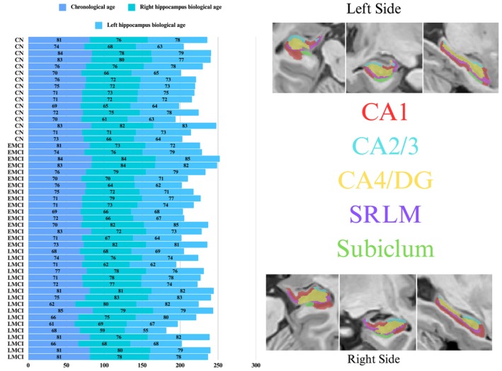
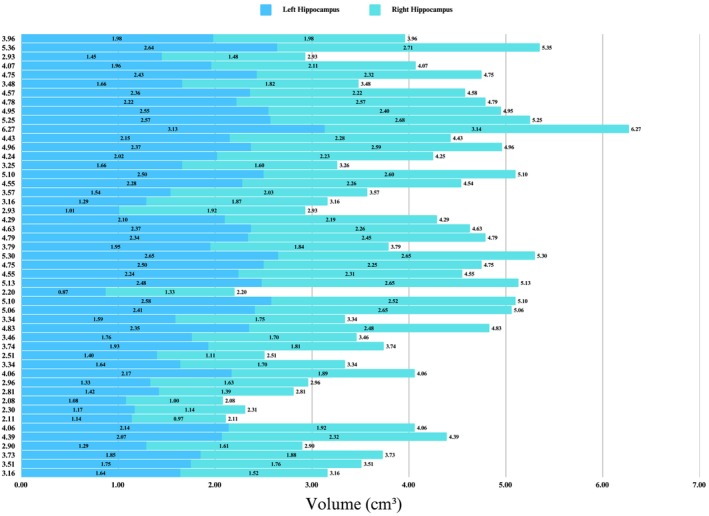
Methods: A cohort of 49 participants, categorized as cognitively normal (CN, n = 16), early MCI (EMCI, n = 16), or late MCI (LMCI, n = 17), underwent comprehensive neuroimaging, neuropsychological, and genetic assessments. High-resolution 3D T1-weighted MRI scans were processed using the volBrain platform and hippocampal subfield segmentation (HIPS) pipeline to quantify hippocampal subfield volumes and estimate biological age. Statistical analyses, including ANCOVA and stepwise regression, were employed to evaluate group differences and identify predictors of hippocampal biological age.
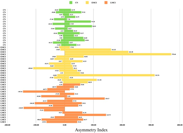
Results: The results revealed significant volumetric reductions in LMCI, particularly within the CA1, CA4/dentate gyrus (DG), and stratum radiatum/lacunosum/moleculare (SRLM) subfields, with pronounced lateralized effects. Clinical and demographic covariates attenuated group differences in biological age, but volumetric adjustments highlighted a significant distinction between EMCI and LMCI, with EMCI exhibiting a higher biological age. Cognitive performance, as measured by the Montreal Cognitive Assessment (MoCA), emerged as a consistent predictor of biological age, while APOE ε4 carrier status was significantly elevated in LMCI patients. Regression analyses identified divergent contributions of CA2/3 (positively associated) and CA4/DG (negatively associated) volumes to biological age, underscoring the subfield-specific pathophysiological mechanisms. Asymmetry indices, although variably expressed across groups, offered limited predictive utility, with CA2/3 and CA4/DG asymmetries modestly influencing biological age.
Conclusion: These findings support the integration of subfield-specific hippocampal volumetry and cognitive assessments in early diagnostic frameworks while highlighting the need for longitudinal studies to elucidate causal pathways linking subfield atrophy, biological aging, and cognitive decline.
Keywords: hippocampal biological age; mild cognitive impairment (MCI); neurodegeneration; neuroimaging; structural MRI.
© 2025 The Author(s). CNS Neuroscience & Therapeutics published by John Wiley & Sons Ltd.
Conflict of interest statement
The authors declare no conflicts of interest.
Figures


Similar articles
-
High‑Resolution T2 MRI Volumetry of Medial Temporal Lobe Subregions Predicts Cognitive Decline Across the Alzheimer's Disease Continuum.Appl Neuropsychol Adult. 2025 Aug 21:1-13. doi: 10.1080/23279095.2025.2546951. Online ahead of print. Appl Neuropsychol Adult. 2025. PMID: 40836826
-
Enhanced Detection of Age-Related and Cognitive Declines Using Automated Hippocampal-To-Ventricle Ratio in Alzheimer's Patients.Hum Brain Mapp. 2025 Aug 1;46(11):e70265. doi: 10.1002/hbm.70265. Hum Brain Mapp. 2025. PMID: 40747973 Free PMC article.
-
Age-Associated Cortical Thinning in Speech Motor Regions Precedes Hippocampal Decline: Implications for Alzheimer's Disease.Hum Brain Mapp. 2025 Aug 1;46(11):e70288. doi: 10.1002/hbm.70288. Hum Brain Mapp. 2025. PMID: 40698900 Free PMC article.
-
Hippocampal subfield volumes in mild cognitive impairment and alzheimer's disease: a systematic review and meta-analysis.Brain Imaging Behav. 2023 Dec;17(6):778-793. doi: 10.1007/s11682-023-00804-3. Epub 2023 Sep 28. Brain Imaging Behav. 2023. PMID: 37768441
-
Unveiling the hippocampal subfield changes across the Alzheimer's disease continuum: a systematic review of neuroimaging studies.Brain Imaging Behav. 2025 Feb;19(1):253-267. doi: 10.1007/s11682-024-00952-0. Epub 2024 Oct 23. Brain Imaging Behav. 2025. PMID: 39443362
References
-
- Liu X., Li H., and Fan Y., “Asymmetric Degeneration of the Hippocampus Predicts Risk of Alzheimer's Disease,” Alzheimer's & Dementia 18, no. S6 (2022): e066271, 10.1002/alz.066271. - DOI
-
- Berron D., Baumeister H., Diers K., et al., “Hippocampal Subregional Thinning Related to Tau Pathology in Early Stages of Alzheimer's Disease,” Alzheimer's & Dementia 18, no. S5 (2022): e067057, 10.1002/alz.067057. - DOI
MeSH terms
Grants and funding
LinkOut - more resources
Full Text Sources
Medical
Miscellaneous
