

Characterization of Vitreous Microbiota Dysbiosis Associated with Proliferative Diabetic Retinopathy
- PMID: 40726501
- PMCID: PMC12301127
- DOI: 10.2147/DMSO.S527069
Characterization of Vitreous Microbiota Dysbiosis Associated with Proliferative Diabetic Retinopathy
Abstract
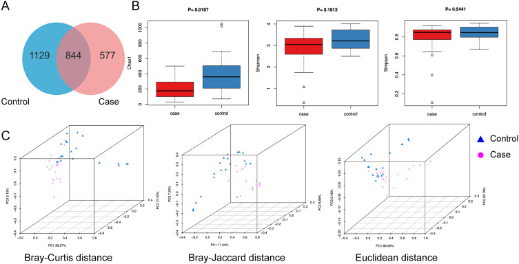
Purpose: Emerging evidence suggests an association between ocular microbiota dysbiosis and ophthalmic diseases; however, the role of the posterior segment microbiome in diabetic retinopathy (DR) remains poorly characterized. In this study, we characterized the vitreous microbiome of patients with proliferative diabetic retinopathy (PDR) and systematically compared its microbial community structure with that of healthy controls.
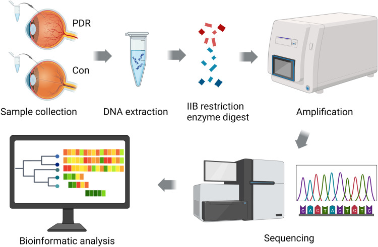
Methods: A cohort of 19 PDR patients with type 2 diabetes mellitus and 19 non-DR controls were enrolled, with vitreous samples obtained through vitrectomy. Vitreous microbial composition was characterized using 2bRAD-M sequencing technology, enabling species-level taxonomic resolution. The comparison of dominant taxa, biomarker analysis and metabolic pathway differences between the two groups were further explored.
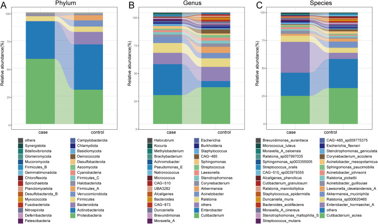
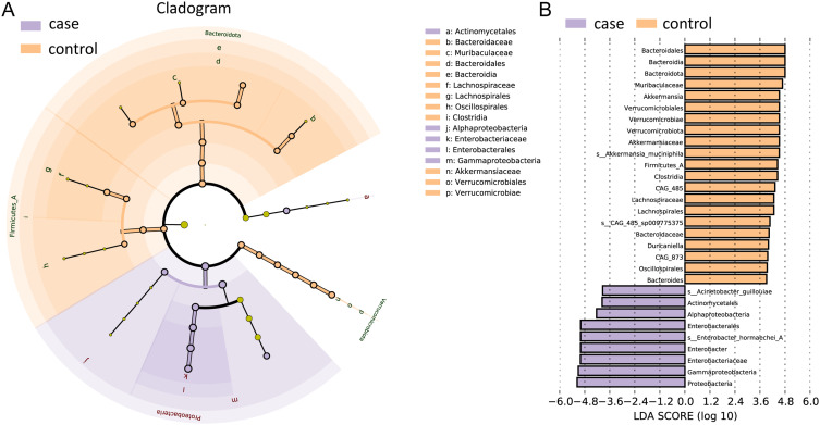
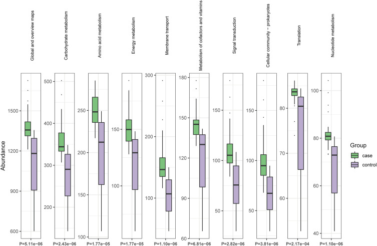
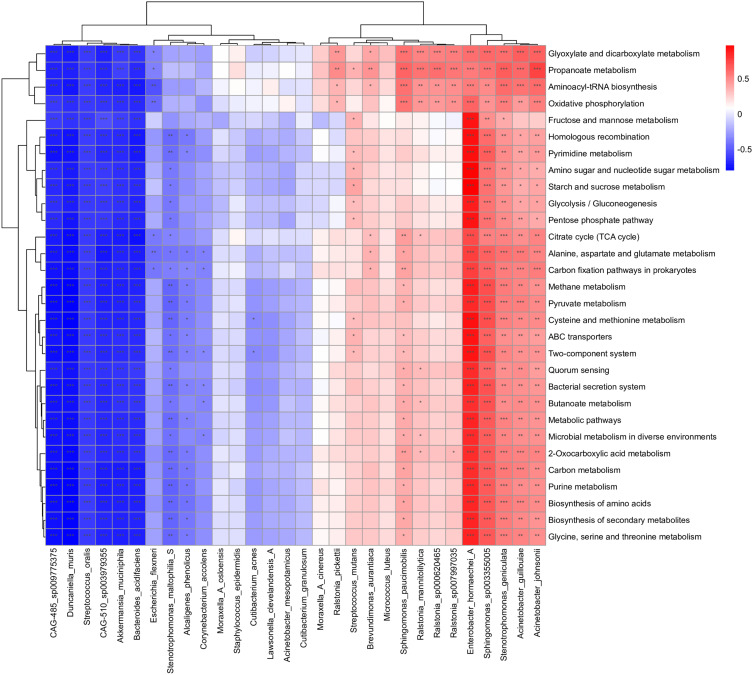
Results: The results of microbiome profiling revealed significant compositional differences in the vitreous core microbiome of PDR patients compared to controls, potentially associated with enhanced activity in membrane transport, nucleotide metabolism and carbohydrate metabolism pathways. LEfSe analysis identified 536 distinctive biomarkers of the two groups. At species level, the PDR group had significantly lower relative abundances of CAG-485_sp009775375, Akkermansia_muciniphila and Bacteroides_acidifaciens, compared with control group.
Conclusion: This is the first study confirming the microbiota in human vitreous fluid samples by 2bRAD-M sequencing. These findings suggest a potential link between vitreous microbial dysbiosis and PDR, offering novel insights for future mechanistic investigations into DR.
Keywords: 2bRAD-M; microbiota; proliferative diabetic retinopathy; vitreous fluid.
© 2025 Song et al.
Conflict of interest statement
The authors report no conflicts of interest in this work.
Figures
Similar articles
-
Anti-vascular endothelial growth factor for proliferative diabetic retinopathy.Cochrane Database Syst Rev. 2014 Nov 24;2014(11):CD008721. doi: 10.1002/14651858.CD008721.pub2. Cochrane Database Syst Rev. 2014. Update in: Cochrane Database Syst Rev. 2023 Mar 20;3:CD008721. doi: 10.1002/14651858.CD008721.pub3. PMID: 25418485 Free PMC article. Updated.
-
Anti-vascular endothelial growth factor for prevention of postoperative vitreous cavity haemorrhage after vitrectomy for proliferative diabetic retinopathy.Cochrane Database Syst Rev. 2015 Aug 7;2015(8):CD008214. doi: 10.1002/14651858.CD008214.pub3. Cochrane Database Syst Rev. 2015. Update in: Cochrane Database Syst Rev. 2023 May 31;5:CD008214. doi: 10.1002/14651858.CD008214.pub4. PMID: 26250103 Free PMC article. Updated.
-
Anti-VEGF drugs compared with laser photocoagulation for the treatment of proliferative diabetic retinopathy: a systematic review and individual participant data meta-analysis.Health Technol Assess. 2025 Apr;29(23):1-75. doi: 10.3310/MJYP6578. Health Technol Assess. 2025. PMID: 40186529 Free PMC article.
-
Anti-VEGF drugs compared with laser photocoagulation for the treatment of diabetic retinopathy: a systematic review and economic analysis.Health Technol Assess. 2025 May;29(23):1-16. doi: 10.3310/KRWP1264. Health Technol Assess. 2025. PMID: 40347224 Free PMC article.
-
Anti-VEGF drugs compared with laser photocoagulation for the treatment of diabetic retinopathy: a systematic review and meta-analysis.Health Technol Assess. 2024 Dec;29(23):1-71. doi: 10.3310/PCGV5709. Health Technol Assess. 2024. PMID: 39673354
References
LinkOut - more resources
Full Text Sources
