

Characterization of Lumpy Skin Disease in Northern Bangladesh: Clinical, Pathological, Biochemical, and Molecular Perspectives
- PMID: 40726513
- PMCID: PMC12303647
- DOI: 10.1155/vmi/4623554
Characterization of Lumpy Skin Disease in Northern Bangladesh: Clinical, Pathological, Biochemical, and Molecular Perspectives
Abstract
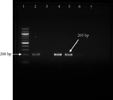
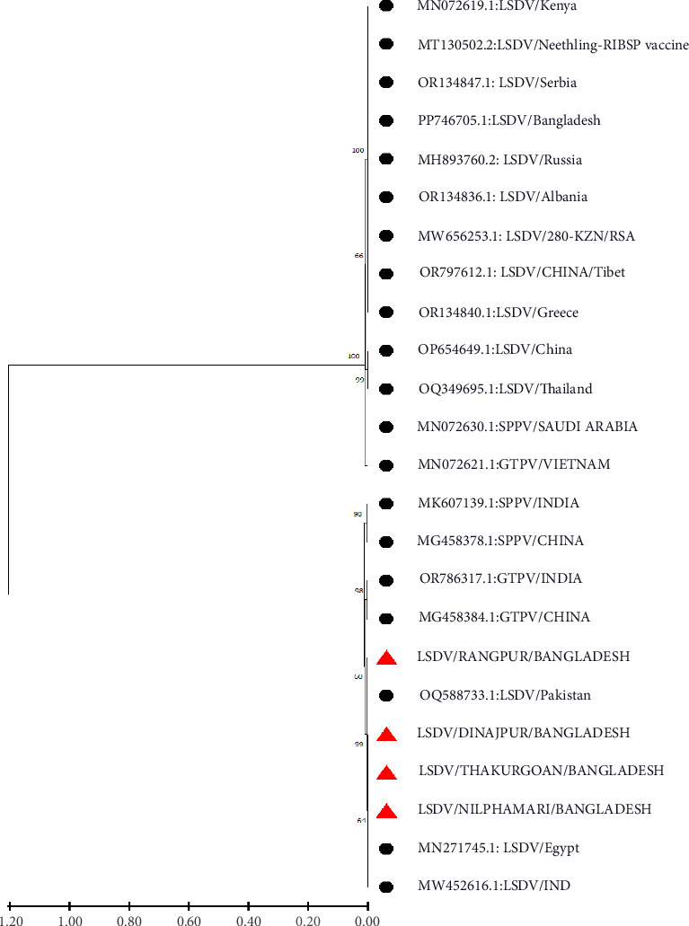
Lumpy skin disease (LSD) is an economically important disease of cattle, considered as a threat to the livestock industry. This study aimed to assess the clinical symptoms, gross pathology and histopathology, serum biochemical values, and molecular characterization of LSD in cattle. Cattle in Dinajpur, Thakurgaon, Nilphamari, and Rangpur affected by LSD were initially diagnosed through clinical signs and gross lesions. Blood samples were then collected for biochemical analysis and molecular detection through PCR, and sequencing was performed to characterize the LSD virus (LSDV). The prevalence of LSD in northern areas of Bangladesh was 33.44%. The recorded clinical signs were high fever; firm, raised skin nodules around the head, neck, and limbs; swelling of the limbs and brisket area; and rough hair coat, nasal discharge, dyspnea, corneal opacity, and severe weakness. Grossly, a well-defined, hard swelling of around 1-3.00 cm in diameter in the skin accompanied enlargement of the nearby lymph nodes, and the nodules are composed of a hard, creamy-gray, or yellow clump of the tissue. Histopathologically, ballooning degeneration of the epidermal cell layers, intracytoplasmic eosinophilic inclusion bodies in distinct dermal and epidermal cells, necrosis with substantial infiltration of inflammatory cells, and congested blood vessels in the dermal layer were noted. While total protein, BUN, serum AST, and ALT were increased significantly (p < 0.05), the serum creatinine, GGT, and total albumin were not changed significantly compared with the healthy cattle. Phylogenetic analysis showed that the circulating isolates from northern part of Bangladesh were found in the same clades with India, Pakistan, and Thailand. Thus, this study provides crucial findings on this emerging disease, including its gross and histopathology, serum biochemical properties, and molecular epidemiology in the northern regions of Bangladesh.
Keywords: clinical signs; gross pathological; histological; phylogenic tree; serum biochemical.
Copyright © 2025 Samiron Roy et al. Veterinary Medicine International published by John Wiley & Sons Ltd.
Conflict of interest statement
The authors declare no conflicts of interest.
Figures


Similar articles
-
Comparative Clinico-Haematological Alterations of Lumpy Skin Disease Affected Cattle at Different Ages and Periods of Disease in Bangladesh.Vet Med Sci. 2025 Sep;11(5):e70549. doi: 10.1002/vms3.70549. Vet Med Sci. 2025. PMID: 40788148 Free PMC article.
-
Molecular characterization of lumpy skin disease virus in the cattle population of District Lower Chitral, Khyber Pakhtunkhwa, Pakistan.Antonie Van Leeuwenhoek. 2025 Aug 3;118(9):124. doi: 10.1007/s10482-025-02137-1. Antonie Van Leeuwenhoek. 2025. PMID: 40754559
-
Molecular Detection, Seroprevalence and Biochemical Analysis of Lumpy Skin Disease Virus.Viruses. 2025 Feb 20;17(3):293. doi: 10.3390/v17030293. Viruses. 2025. PMID: 40143224 Free PMC article.
-
Signs and symptoms to determine if a patient presenting in primary care or hospital outpatient settings has COVID-19.Cochrane Database Syst Rev. 2022 May 20;5(5):CD013665. doi: 10.1002/14651858.CD013665.pub3. Cochrane Database Syst Rev. 2022. PMID: 35593186 Free PMC article.
-
Systemic pharmacological treatments for chronic plaque psoriasis: a network meta-analysis.Cochrane Database Syst Rev. 2021 Apr 19;4(4):CD011535. doi: 10.1002/14651858.CD011535.pub4. Cochrane Database Syst Rev. 2021. Update in: Cochrane Database Syst Rev. 2022 May 23;5:CD011535. doi: 10.1002/14651858.CD011535.pub5. PMID: 33871055 Free PMC article. Updated.
References
-
- Lubinga J. C., Tuppurainen E. S. M., Mahlare R., Coetzer J. A. W., Stoltsz W. H., Venter E. H. Evidence of Transstadial and Mechanical Transmission of Lumpy Skin Disease Virus by Amblyomma hebraeum Ticks. Transboundary and Emerging Diseases . 2015;62(2):174–182. doi: 10.1111/tbed.12102. - DOI - PubMed
-
- Abera Z. Survey on Distribution, Associated Factors of Lumpy Skin Disease Occurrence and its Vaccine Efficacy in Selected Districts of East Wollega Zone, Western Oromiya. Biomedical Journal of Scientific & Technical Research . 2019;13(2) doi: 10.26717/BJSTR.2019.13.002385. - DOI
LinkOut - more resources
Full Text Sources
Miscellaneous
