. 2025 Jun 26;11(7):1207-1217.
doi: 10.1021/acscentsci.5c00959. eCollection 2025 Jul 23.

DCAF16-Based Covalent Degradative Handles for the Modular Design of Degraders

Affiliations

DCAF16-Based Covalent Degradative Handles for the Modular Design of Degraders

Lauren M Orr et al. ACS Cent Sci. .

Abstract

While targeted protein degradation is a powerful strategy for eliminating disease-causing proteins, the rational design of monovalent or molecular glue degraders remains challenging. In this study, we generated a library of BET-domain inhibitor JQ1 analogs bearing elaborated electrophilic handles to identify permissive covalent degradative handles and E3 ligase pairs. We identified an elaborated fumaramide handle that, when appended onto JQ1, led to the proteasome-dependent degradation of BRD4. We revealed that the E3 ubiquitin ligase CUL4DCAF16a common E3 ligase target of electrophilic degraderswas responsible for BRD4 loss by covalently targeting C173 on DCAF16. While this original fumaramide handle was not permissive to the degradation of other neo-substrates, a truncated version of this handle attached to JQ1 was still capable of degrading BRD4, now through targeting both C173 and C178. This truncated fumaramide handle, when appended to various protein targeting ligands, was also more permissive in degrading other neo-substrates, including CDK4/6, SMARCA2/4, the androgen receptor (AR), as well as the undruggable AR truncation variant AR-V7. We have identified a unique DCAF16-targeting covalent degradative handle that can be transplanted across several protein-targeting ligands to induce the degradation of their respective targets for the modular design of monovalent or bifunctional degraders.


Figures

1
1
Screening of JQ1 analogs bearing electrophilic handles for BRD4 degradation. (a) Structures of JQ1 analogs bearing electrophilic handles. (b) Screening JQ1 electrophilic library in HEK293T cells for BRD4 degradation. HEK293T cells were treated with DMSO vehicle or compounds (1 μM) for 24 h and BRD4 and loading control actin levels were assessed by SDS/PAGE and Western blotting. (c) Structure of best hit HRG038 with the elaborated electrophilic handle in red. (d) Quantification of the experiment is described in (b). Blot in (b) represents n = 3 biologically independent replicates per group. Data in (d) show individual replicates and average ± sem from n = 3 biologically independent replicates per group. Significance expressed as *p < 0.05 compared to vehicle-treated controls.
2
2
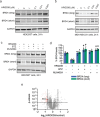
Characterization of HRG038 as a BRD4 degrader. (a, b) Dose–response of BRD4 degradation with HRG038 treatment. HEK293T cells (a) or MDA-MB-231 cells (b) were treated with DMSO vehicle or HRG038 for 24 h, after which BRD4 and loading control GAPDH levels were assessed by SDS/PAGE and Western blotting. (c) Proteasome-dependence of BRD4 degradation by HRG038. HEK293T cells were pretreated with DMSO vehicle, BTZ (1 μM), or MLN4924 (1 μM) for 1 h prior to treatment of cells with DMSO or HRG038 (100 nM) for 24 h after which BRD4 and loading control GAPDH levels were assessed by SDS/PAGE and Western blotting. (d) Quantification of data from the experiment described in (c). (e) Quantitative tandem mass tagging (TMT)-based proteomic profiling of HRG038 in MDA-MB-231 cells. MDA-MB-231 cells were treated with DMSO vehicle or HRG038 (1 μM) for 24 h. Shown in red are proteins significantly lowered in levels with log2 < 0.6 with p < 0.01 with BRD4 highlighted. Full proteomics data can be found in . Blots in (a-c) are representative of n = 3 biologically independent replicates per group. Bar graph in (d) shows individual replicate values and average ± sem from n = 3 biologically independent replicates per group. Data in (e) are from n = 3 biologically independent replicates per group. Significance in (d) shown as *p < 0.01 compared to vehicle-treated controls and #p < 0.05 compared to HRG038-treated groups.
3
3
Functional CRISPR screen with UBAL library to identify E3 ligase responsible for HRG038-mediated BRD4 degradation. (a) A schematic for the CRISPR-Cas9 screen design. We performed parallel screens with the BRD4-GFP reporter cells expressing Cas9 treated with vehicle or HRG038 (in duplicate). (b,c) Volcano plots of the screen data for the vehicle (b) and HRG038 (c). The y-axis is a confidence score, and the x-axis indicates the maximum effect (phenotype) size, with more positive values indicating an enrichment of the sgRNAs in the high GFP population, while more negative values reflecting an enrichment in the low GFP population. The confidence score is based on the Cas9 High-Throughput Maximum Likelihood Estimator (casTLE) score. (d) Comparative analyses of the confidence scores in (b) and (c). This analysis highlights hits that are part of the endogenous pathway (e.g., SPOP) versus hits that are selective to the degrader molecule (e.g., hits that sit on the y-axis, DCAF16/DDB1). (e) Histograms showing the enrichment of multiple sgRNAs per gene within cells with high levels of BRD4-GFP (i.e., degradation inhibited by gene KO). Functional CRISPR screen data can be found in .
4
4
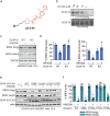
Characterizing CUL4DCAF16 as the responsible target for HRG038-mediated BRD4 degradation. (a) Alkyne-functionalized probe of degradative handle, LO-3-44. (b) LO-3-44 labeling of pure human CUL4DCAF16. The CUL4DCAF16-DDB1-DDA1 complex was labeled with LO-3-44 for 1 h, after which probe-labeled proteins were conjugated with an azide-functionalized rhodamine by copper-mediated azide–alkyne cycloaddition (CuAAC). Proteins were separated by SDS/PAGE and visualized by in-gel fluorescence or loading was assessed by silver staining. (c) BRD4 degradation in CUL4DCAF16 WT and KO cells. CUL4DCAF16 WT and KO HEK293T cells were treated with DMSO vehicle or HRG038 (100 nM) for 24 h and BRD4 and loading control GAPDH levels were assessed by SDS/PAGE and Western blotting. (d) Quantification for the experiment described in (c). (e) HRG038-mediated BRD4 degradation in cells expressing WT and mutant CUL4DCAF16. CUL4DCAF16 KO HEK293T cells expressing either empty vector, FLAG-CUL4DCAF16 WT, C58S, C119S, C173S, or C178S were treated with DMSO vehicle or HRG038 (100 nM) for 24 h and BRD4 and loading control actin levels were assessed by SDS/PAGE and Western blotting. (f) Quantification for the experiment described in (d). Gels and blots in (b,c,e) are representative of n = 3 biologically independent replicates/group, of which individual replicates and average ± sem are shown in (d,f). Significance is expressed as *p < 0.05 compared to vehicle-treated WT controls in (d) or respective vehicle-treated groups (f) and #p < 0.05 compared to CUL4DCAF16 WT cells treated with HRG038 in (d) or CUL4DCAF16 KO cells expressing FLAG-CUL4DCAF16 WT treated with HRG038 (f).
5
5
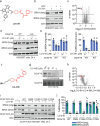
Assessing the degradative potential of truncated fumaramide handle. (a) Structure of LO-3-61, a JQ1 analog bearing a truncated fumaramide handle in red. (b) Proteasome-dependence of BRD4 degradation by HRG038. HEK293T cells were pretreated with DMSO vehicle, BTZ (1 μM) for 1 h prior to treatment of cells with DMSO or LO-3-61 (100 nM) for 24 h after which BRD4 and loading control GAPDH levels were assessed by SDS/PAGE and Western blotting. (c) Quantitative tandem mass tagging (TMT)-based proteomic profiling of LO-3-61 in K562 cells. K562 cells were treated with DMSO vehicle or LO-3–61 (100 nM) for 24 h. Shown in red are proteins significantly lowered in levels with log2 < 0.6 with p < 0.01 with BRD4 highlighted. Full proteomics data can be found in . (d) BRD4 degradation in CUL4DCAF16 WT and KO cells. CUL4DCAF16 WT and KO HEK293T cells were treated with DMSO vehicle or LO-3–61 for 24 h, and BRD4 and loading control actin levels were assessed by SDS/PAGE and Western blotting. (e) Quantification of the experiment described in (d). (f) The structure of LO-4-06, an alkyne-functionalized probe based on the truncated fumaramide handle shown in red. (g) LO-4–06 labeling of pure human CUL4DCAF16. The CUL4DCAF16-DDB1-DDA1 complex was labeled with LO-4-06 for 1h, after which probe-labeled proteins were conjugated with an azide-functionalized rhodamine by copper-mediated azide–alkyne cycloaddition (CuAAC). Proteins were separated by SDS/PAGE and visualized by in-gel fluorescence or loading was assessed by silver staining. (h) Pulldown chemoproteomics profiling with the probe. HEK293T cell lysates were pretreated with DMSO vehicle or LO-3-61 (50 μM) 1 h prior to treatment with LO-4-06 probe (10 μM). Probe-modified proteins were appended with azide-functionalized biotin by copper-mediated azide–alkyne cycloaddition (CuAAC), after which probe-modified proteins were avidin-enriched, tryptically digested, and quantitatively analyzed by TMT-based proteomics. Shown in red are proteins in which LO-4–06 labeling was significantly out-competed by LO-3-61, with DCAF16 highlighted. (i) LO-3–61-mediated BRD4 degradation in cells expressing WT and mutant CUL4DCAF16. CUL4DCAF16 KO HEK293T cells expressing either empty vector, FLAG-CUL4DCAF16 WT, C58S, C119S, C173S, or C178S were treated with DMSO vehicle or LO-3–61 (100 nM) for 24 h and BRD4 and loading control actin levels were assessed by SDS/PAGE and Western blotting. (j) Quantification for the experiment described in (i). Gels and blots in (b,d,g,i) are representative of n = 3 biologically independent replicates per group with individual replicates and average ± sem are shown in (e,j). Data in (c,h) are from n = 3 biologically independent replicates per group and proteomics data can be found in and , respectively.
6
6
Model of CUL4DCAF16-DDB1-DDA1 complex showing C173, C177–C179, and C58. The model is based on a structure previously solved of DCAF16-DDB1-DDA1 in complex with BRD4 in the presence of an electrophilic BRD4 molecular glue degrader MMH2 (PDB: 8g46) that did not include the disordered loop containing C173. We have removed MMH2 and BRD4. To add the loop containing C173 not resolved in the reported structure, the PyMol builder function was used to add the residues to the model structure, and the loop was then fit to a potential conformation via homology modeling with ModLoop. , The structure is adapted from PDB: 8g46 (ref ). Rights to use and adapt this structure can be found in http://creativecommons.org/licenses/by/4.0/.
7
7
Permissiveness of truncated fumaramide handle in degrading multiple neo-substrates. (a) Structure of LO-3-63, a CDK4/6 inhibitor ribociclib appended to truncated fumaramide handle in red. (b) CDK4/6 degradation in HEK293T cells. HEK293T cells were treated with DMSO vehicle or LO-3-63 for 24 h after which CDK4/6 and loading control actin levels were assessed by SDS/PAGE and Western blotting. (c) CDK4/6 degradation CUL4DCAF16 WT and KO cells. CUL4DCAF16 WT and KO HEK293T cells were treated with DMSO vehicle or LO-3-63 for 12 h after which CDK4/6 and loading control actin levels were assessed by SDS/PAGE and Western blotting. (d) Structure of LO-3-62, a SMARCA2/4 inhibitor linked to truncated fumaramide handle in red. (e) SMARCA2/4 degradation in MV-4-11 cells. MV-4-11 cells were treated with DMSO vehicle or LO-3-62 for 24 h after which SMARCA2/4 and loading control actin levels were assessed by SDS/PAGE and Western blotting. (f) Structure of LO-4-25, a AR DNA binding domain ligand linked via a linker to the truncated fumaramide handle in red. (g) AR and AR-V7 degradation in 22Rv1 cells. 22Rv1 cells were treated with DMSO vehicle or LO-4-25 for 24 h after which AR and AR-V7 levels were assessed by SDS/PAGE and Western blotting. (h) DCAF16 mRNA levels. 22Rv1 cells were transiently transfected with siControl (−) or siDCAF16 (+) oligonucleotides for 48 h after which DCAF16 mRNA levels were assessed by qPCR. (i) AR and AR-V7 degradation in siControl and siDCAF16 22Rv1 cells. 22Rv1 siControl or siDCAF16 cells were treated with DMSO vehicle or LO-4–25 for 24 h and AR, AR-V7, and loading control actin levels were assessed by SDS/PAGE and Western blotting. (j) Quantification of the experiment described in (i). Blots in (b,c,e,g,i) are representative of n = 3 biologically independent replicates/group. Bar graphs in (h,j) show individual replicate values and average ± SEM. Significance expressed as *p < 0,05 compared to siControl in (h) or to vehicle-treated siControl groups in (j) and #p < 0.05 compared to LO-4-25-treated siControl groups in (j).

References

    1. Zhang X.. et al. DCAF11 Supports Targeted Protein Degradation by Electrophilic Proteolysis-Targeting Chimeras. J. Am. Chem. Soc. 2021;143:5141–5149. doi: 10.1021/jacs.1c00990. - DOI - PMC - PubMed
    1. Campos M. A.. et al. Discovery of DCAF16 Binders for Targeted Protein Degradation. ACS Chem. Biol. 2025;20:479–488. doi: 10.1021/acschembio.4c00799. - DOI - PMC - PubMed
    1. Zhang X., Crowley V. M., Wucherpfennig T. G., Dix M. M., Cravatt B. F.. Electrophilic PROTACs that degrade nuclear proteins by engaging DCAF16. Nat. Chem. Biol. 2019;15:737–746. doi: 10.1038/s41589-019-0279-5. - DOI - PMC - PubMed
    1. Hassan M. M.. et al. Exploration of the tunability of BRD4 degradation by DCAF16 trans-labelling covalent glues. Eur. J. Med. Chem. 2024;279:116904. doi: 10.1016/j.ejmech.2024.116904. - DOI - PMC - PubMed
    1. Lim M.. et al. DCAF16-Based Covalent Handle for the Rational Design of Monovalent Degraders. ACS Cent. Sci. 2024;10:1318–1331. doi: 10.1021/acscentsci.4c00286. - DOI - PMC - PubMed

LinkOut - more resources