. 2025 Jul 16:33:102106.
doi: 10.1016/j.mtbio.2025.102106. eCollection 2025 Aug.

Tannic acid-iron stabilized probiotic silver nano hybrids: Multi-target gut microbiota modulation and intestinal barrier restoration

Affiliations

Tannic acid-iron stabilized probiotic silver nano hybrids: Multi-target gut microbiota modulation and intestinal barrier restoration

Saisai Gong et al. Mater Today Bio. .

Abstract

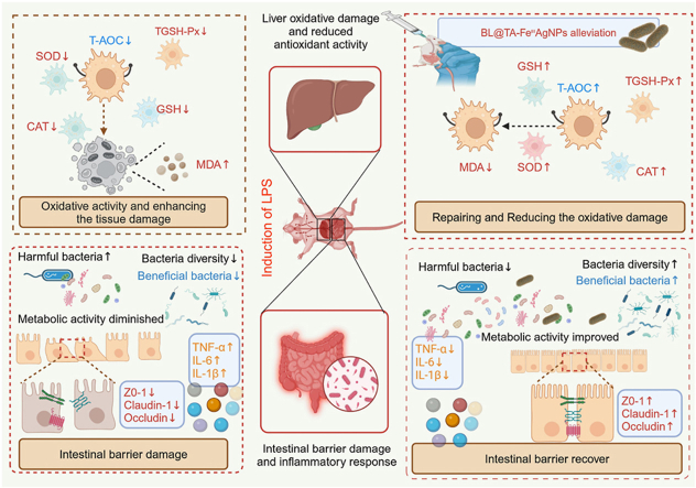
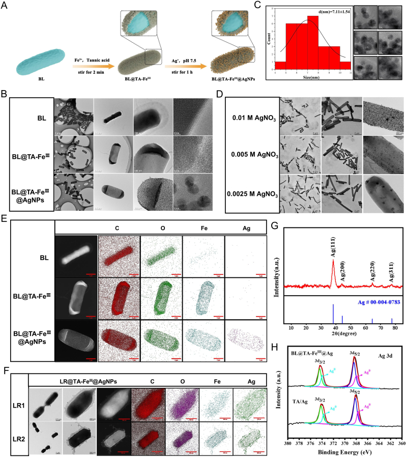
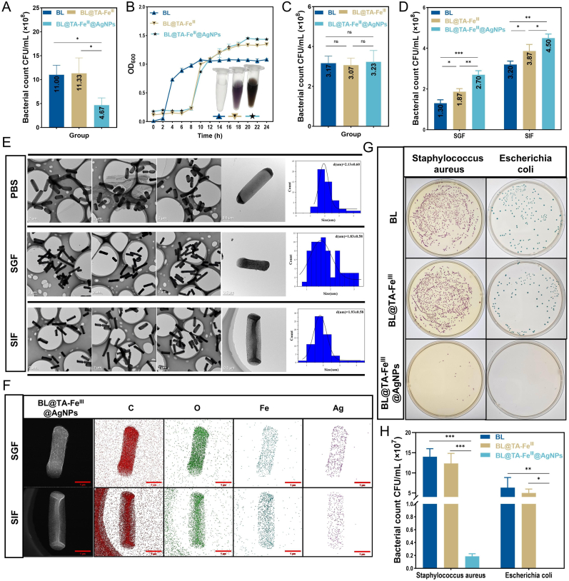
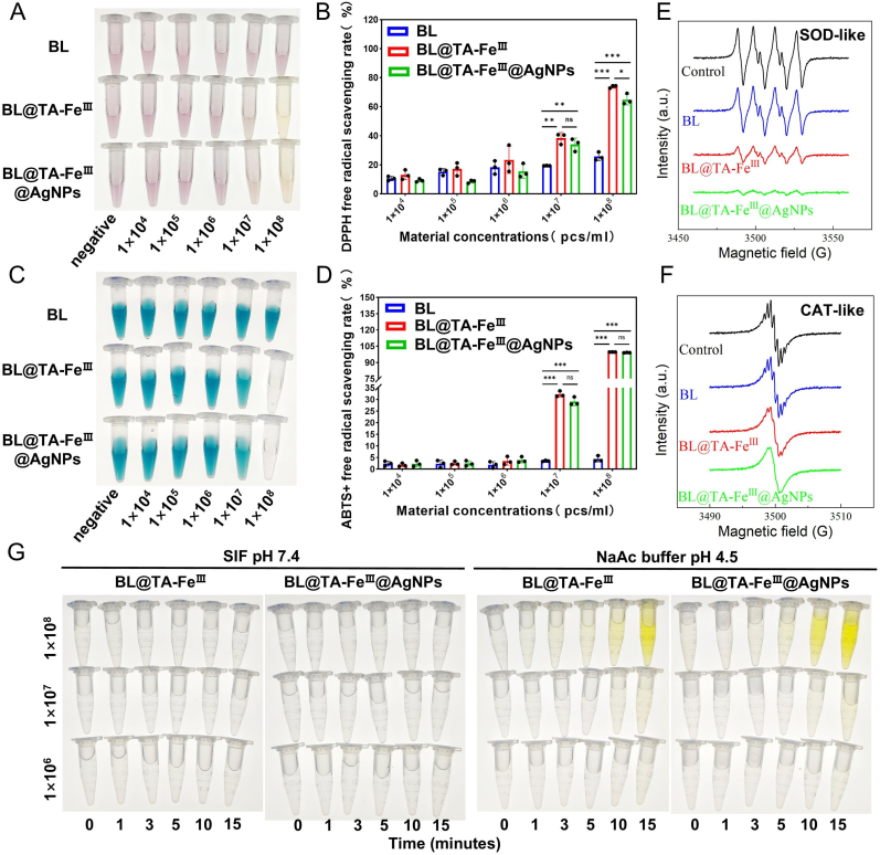
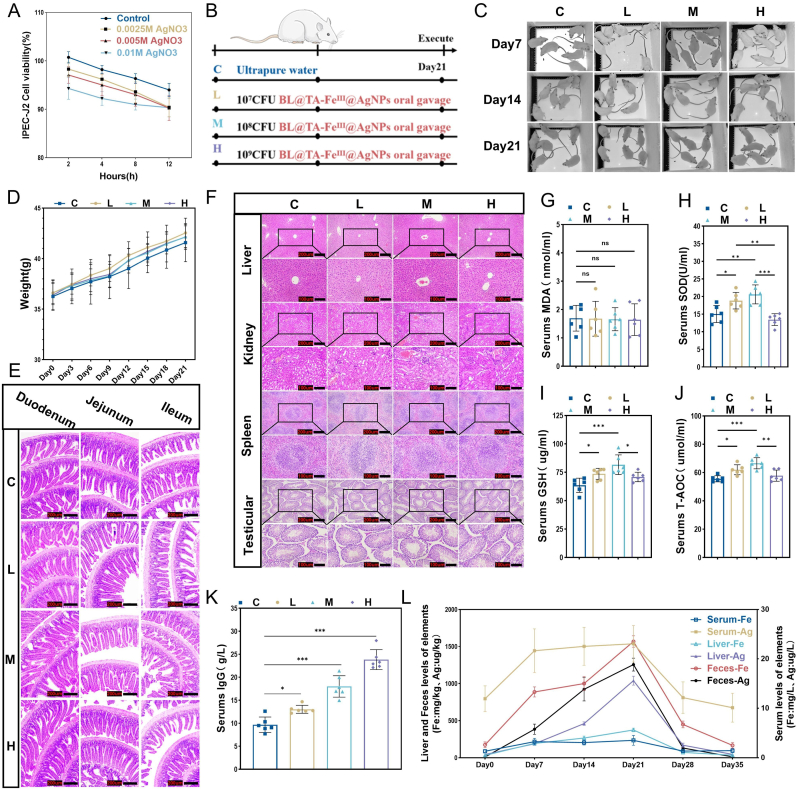
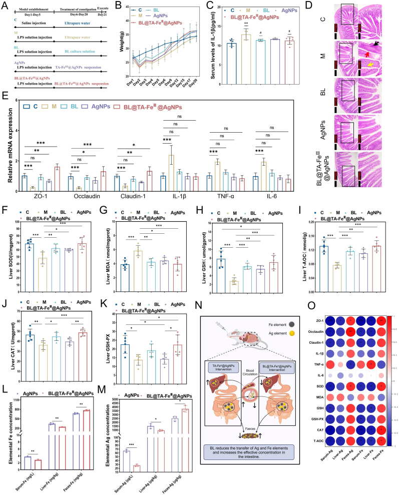
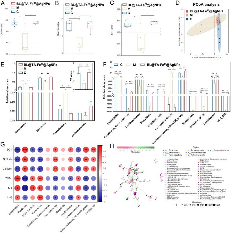
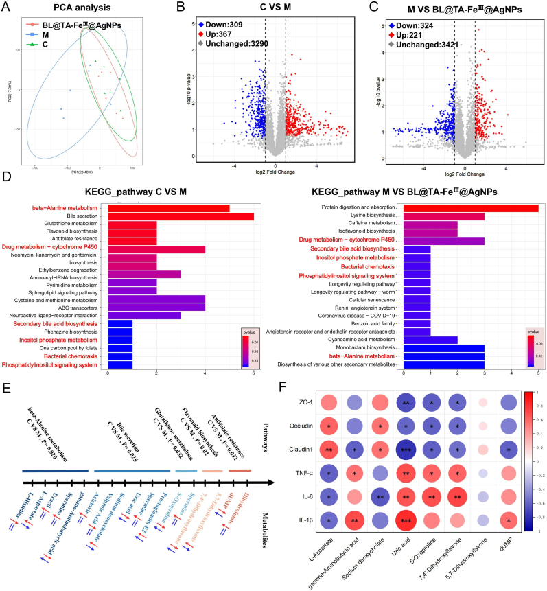
The limited efficacy and adverse effect profile of current pharmacological treatments for intestinal inflammation underscore the need for modalities that preserve gut microbiota balance while attenuating inflammation. The aim of this study was to develop and evaluate a BL@TA-FeIII@AgNPs system with a view to provide synergistic efficacy against intestinal injury. This research introduces an innovative hybrid bio nanocomposite, BL@TA-FeIII@AgNPs, comprising viable Bacillus licheniformis coated with a tannic-acid/FeIII coordination layer that nucleates and anchors 7 ± 1.5 nm silver nanoparticles. Characterization of this composite material was performed using TEM, EDS, XRD, and XPS. Functional assays included probiotic viability, tolerance to simulated gastric and intestinal fluids, and bactericidal activity against Escherichia coli and Staphylococcus aureus. In-depth safety evaluations were carried out using both cell cultures and a mouse model. Therapeutic effects in an acute LPS-endotoxemia mouse model were analyzed by 16S rRNA gene amplicon sequencing and untargeted LC-MS/MS metabolomics of cecal contents. Characterization confirmed structural integrity, colloidal stability in physiological media, and low cytotoxicity (IC50 > 100 μg Ag mL-1). BL@TA-FeIII@AgNPs restored transepithelial electrical resistance, lowered malondialdehyde levels, and reshaped microbiota composition and metabolite networks relative to LPS controls. Restoration of the Firmicutes: Bacteroidetes ratio and elevated short chain fatty acid concentrations support BL@TA-FeIII@AgNPs as a promising adjunctive strategy for acute endotoxin-induced intestinal injury.

Keywords: Bio-nanocomposites; Gut microbiota; Intestinal inflammation; Nanomedicine; Probiotics; Silver nanoparticles.


Conflict of interest statement

The authors declare that they have no known competing financial interests or personal relationships that could have appeared to influence the work reported in this paper.

Figures

Image 1
Graphical abstract
Fig. 1
Fig. 1
The construction and morphological characterization of BL@TA-FeIII@AgNPs. (A) Schematic of the synthesis of BL@TA-FeIII@AgNPs. (B) TEM images of BL, BL@TA-FeIII, BL@TA-FeIII@AgNPs. (C) Particle size distribution of nanoparticles on the surface of BL@TA-FeIII@AgNPs. (D) TEM images of BL@TA-FeIII@AgNPs prepared with different concentrations of AgNO3. (E) EDS elemental analysis patterns of BL, BL@TA-FeIII, BL@TA-FeIII@AgNPs. (F) TEM image and EDS element map of LR@ TA-FeIII@AgNPs (composite material based on Lactobacillus reuteri). (G) XRD analysis of BL@TA-FeIII@AgNPs. (H) XPS patterns of Ag 3d of BL@TA-FeIII@AgNPs and TA-FeIII@AgNPs.
Fig. 2
Fig. 2
The adaptability of the TA-FeIII@AgNPs shell to the environment and its impact on the activity of BL. (A) Effect of TA-FeIII and TA-FeIII@AgNPs shells on the number of BL survivors. (B) Effect of TA-FeIII and TA-FeIII@AgNPs shells on BL growth curves. (C) Effect of TA-FeIII and TA-FeIII@AgNPs shells on the number of BL surviving at plateau stage. (D) Effect of TA-FeIII and TA-FeIII@AgNPs shells on BL resistance to gastrointestinal fluid. (E) TEM images and bacterial length distribution of BL@TA-FeIII@AgNPs after treatment with PBS, SGF, and SIF. (F) EDS elemental mapping scans of BL@TA-FeIII@AgNPs after SGF and SIF treatment. (G, H) Bactericidal effect and viable counts of BL, BL@TA-FeIII, and BL@TA-FeIII@AgNPs. Significant variations were denoted by ∗ (p < 0.05), ∗∗ (p < 0.01), or ∗∗∗ (p < 0.001).
Fig. 3
Fig. 3
Multi-Enzyme mimetic antioxidant activity of BL@TA-FeIII@AgNPs shells. (A,B) Ability of BL, BL@TA-FeIII and BL@TA-FeIII@AgNPs to scavenge DPPH free radicals at different concentrations. (C, D) Ability of BL, BL@TA-FeIII and BL@TA-FeIII@AgNPs to scavenge ABTS free radicals at different concentrations. (E) EPR signaling profiles of superoxide radical adducts generated by BL, BL@TA-FeIII and BL@TA-FeIII@AgNPs after incubation, reflecting their SOD-like activities. (F) EPR signaling profiles of BL, BL@TA-FeIII and BL@TA-FeIII@AgNPs cultured in H2O2 solution after H2O2 removal, reflecting their CAT-like activity. (G) Intestinal or in vitro POD-like activities of BL@TA-FeIII and BL@TA-FeIII@AgNPs at different concentrations of BL@TA-FeIII and BL@TA-FeIII@AgNPs. Significant variations were denoted by ∗ (p < 0.05), ∗∗ (p < 0.01), or ∗∗∗ (p < 0.001).
Fig. 4
Fig. 4
The biocompatibility assessment of BL@TA-FeIII@AgNPs. (A) Effect of BL@TA-FeIII@AgNPs prepared with different concentrations of AgNO3 on the proliferation of IPEC-J2 cells. (B) Intervention procedures of different doses of BL@TA-FeIII@AgNPs on mice. (C) Effects of different doses of BL@TA-FeIII@AgNPs on the mental state of mice as reflected by fur condition. (D) Effects of different doses of BL@TA-FeIII@AgNPs on body weight of mice (n = 10). (E) Effects of different doses of BL@TA-FeIII@AgNPs on the intestinal tissue structure of mice. (F) Effects of different doses of BL@TA-FeIII@AgNPs on the tissue structure of the liver, kidneys, spleen, and testes of mice. Effects of different doses of BL@TA-FeIII@AgNPs on serum antioxidant assessment-related indexes MDA (G), SOD (H), GSH (I), T-AOC (J) in mice of different doses of BL@TA-FeIII@AgNPs on the intestinal tissue structure of mice (n = 6). (K) Effects of different doses of BL@TA-FeIII@AgNPs on serum IgG levels in mice of different doses of BL@TA-FeIII@AgNPs on the intestinal tissue structure of mice (n = 6). (L) Metabolism of elements Fe, Ag in BL@TA-FeIII@AgNPs in feces, serum and liver of different doses of BL@TA-FeIII@AgNPs on the intestinal tissue structure of mice (n = 3). Significant variations were denoted by ∗ (p < 0.05), ∗∗ (p < 0.01), or ∗∗∗ (p < 0.001).
Fig. 5
Fig. 5
BL@TA-FeIII@AgNPs improve LPS-induced intestinal barrier damage and hepatic antioxidant capacity decline. (A) LPS exposure procedures and interventions. (B) Body weight (n = 12).(C)Serum IL-1β level (n = 6). (D) Pathologic observation of jejunum. Histological assessment of intestinal tissue showing abnormal overall structure with regular villi arrangement (yellow arrow indicates necrotic mucosal epithelial cells), increased number of goblet cells (red arrow), and mild inflammatory cell infiltration (black arrow). (E) Jejunal tight junction-related gene and inflammatory factor expression levels (n = 6). Assessment of oxidative and antioxidant substances in liver tissue (n = 6). (F) SOD; (G) MDA; (H) GSH; (I) T-AOC; (J) CAT; (K) GSH-Px. Levels of elemental Fe (L) and Ag (M) in serum, liver and feces (n = 3). (N) Roadmap for the transfer of the elements Fe and Ag. (O) Heatmap of the correlation between intestinal barrier genes, pro-inflammatory factors, and liver antioxidant indicators with the contents of Ag and Fe elements. Significant variations were denoted by ∗ (p < 0.05), ∗∗ (p < 0.01), or ∗∗∗ (p < 0.001). (For interpretation of the references to colour in this figure legend, the reader is referred to the Web version of this article.)
Fig. 6
Fig. 6
BL@TA-FeIII@AgNPs ameliorate LPS-induced gut bacterial dysbiosis (n = 5). α-diversity analysis, Chao1 index (A), Shannon index (B), ACE index (C).β-diversity analysis, PCoA score plot (D). (E) phylum-level differential bacterial analysis. (F) Genus level differential bacteria analysis. (G) Correlation of differential bacteria with gut barrier genes and pro-inflammatory factors. (H) Inter-regulatory network relationships of gut microbiota. Significant variations were denoted by ∗ (p < 0.05), ∗∗ (p < 0.01), or ∗∗∗ (p < 0.001).
Fig. 7
Fig. 7
BL@TA-FeIII@AgNPs ameliorates LPS-induced gut metabolic disorders (n = 6). (A)PCA analysis. (B)Differential metabolite expression volcano plots of group C vs. group M. (C)Differential metabolite expression volcano plots of group C and BL@TA-FeIII@AgNPs treated group. (D)Differential metabolite KEGG functional enrichment analysis. (E)Analysis of differential metabolic pathways and differential metabolites under the common influence,C vs M (red arrow), M vs BL@TA-FeIII@AgNPs (blue arrow). (F)Correlation analysis of differential metabolites with potentially important functions with gut barrier genes and pro-inflammatory factors. Significant variations were denoted by ∗ (p < 0.05), ∗∗ (p < 0.01), or ∗∗∗ (p < 0.001). (For interpretation of the references to colour in this figure legend, the reader is referred to the Web version of this article.)

Similar articles

References

    1. Winter S.E., Keestra A.M., Tsolis R.M., Baumler A.J. The blessings and curses of intestinal inflammation. Cell Host Microbe. 2010;8:36–43. doi: 10.1016/j.chom.2010.06.003. - DOI - PMC - PubMed
    1. Gupta S., Sharma A.K., Shastri V., Madhu M.K., Sharma V.K. Prediction of anti-inflammatory proteins/peptides: an insilico approach. J. Transl. Med. 2017;15:7. doi: 10.1186/s12967-016-1103-6. - DOI - PMC - PubMed
    1. Kangwan N., Kongkarnka S., Boonkerd N., Unban K., Shetty K., Khanongnuch C. Protective effect of probiotics isolated from traditional fermented tea leaves (miang) from northern Thailand and role of synbiotics in ameliorating experimental ulcerative colitis in mice. Nutrients. 2022;14 doi: 10.3390/nu14010227. - DOI - PMC - PubMed
    1. Ransford R.A., Langman M.J. Sulphasalazine and mesalazine: serious adverse reactions re-evaluated on the basis of suspected adverse reaction reports to the committee on safety of medicines. Gut. 2002;51:536–539. doi: 10.1136/gut.51.4.536. - DOI - PMC - PubMed
    1. Schwartz D.J., Langdon A.E., Dantas G. Understanding the impact of antibiotic perturbation on the human microbiome. Genome Med. 2020;12:82. doi: 10.1186/s13073-020-00782-x. - DOI - PMC - PubMed

LinkOut - more resources