. 2025 Jul 14:27:3167-3180.
doi: 10.1016/j.csbj.2025.07.027. eCollection 2025.

Artificial intelligence insight on structural basis and small molecule binding niches of NMDA receptor

Affiliations

Artificial intelligence insight on structural basis and small molecule binding niches of NMDA receptor

Yunsheng Liu et al. Comput Struct Biotechnol J. .

Abstract

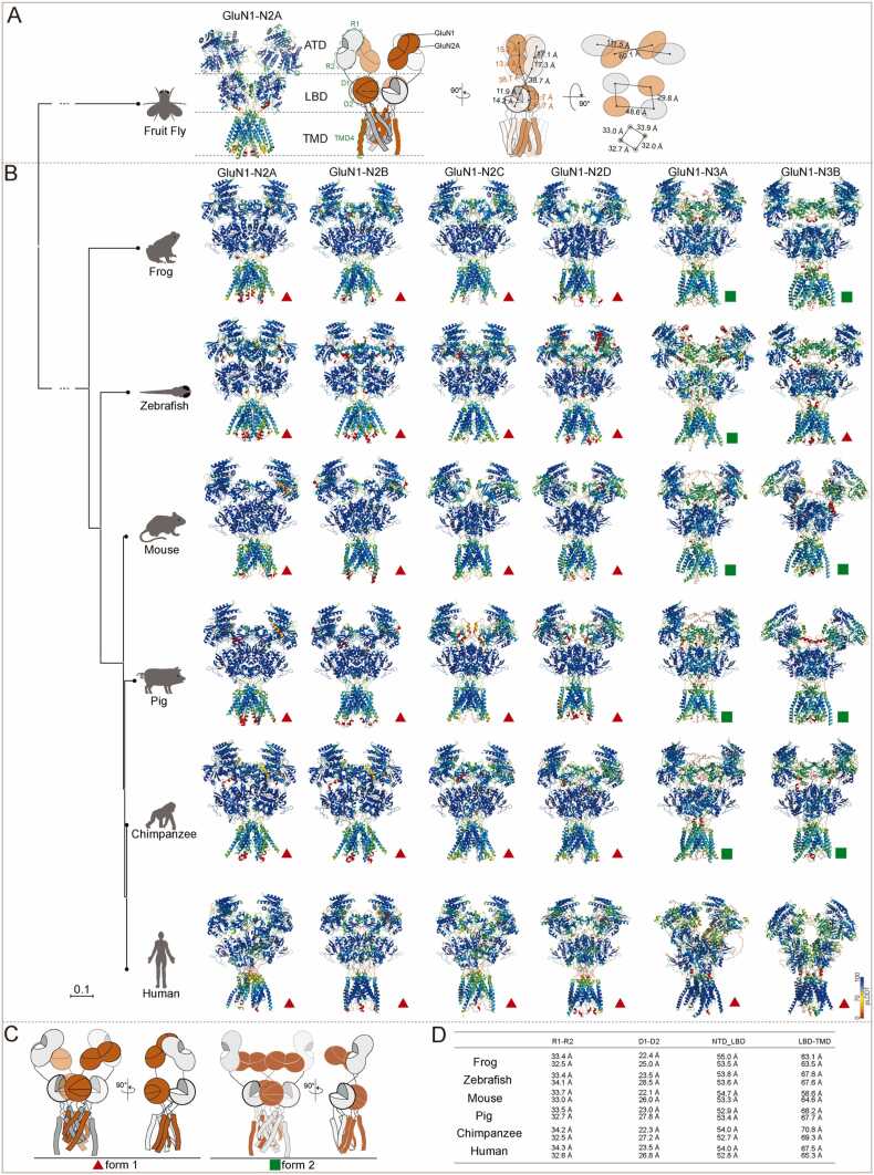
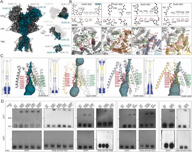
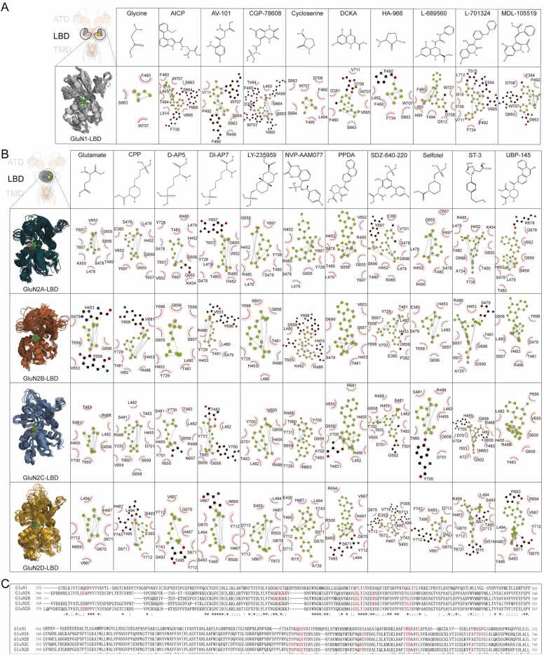
NMDA receptors are critical to neuronal activity and play essential roles in synaptic transmission, learning, and memory. Despite significant advances in X-ray crystallography and cryo-electron microscopy (cryo-EM), the structural diversity of NMDA receptors across species and the variations among receptor subtypes within the same species remain insufficiently explored. Additionally, several key small molecule binding sites, such as those for agonists, antagonists, and allosteric modulators, have not been fully characterized. In this study, we utilized state-of-the-art artificial intelligence algorithms to model NMDA receptors across multiple species and found that they all adopted a bouquet-like dimer-of-dimer structure. By comparing these models with cryo-EM resolved structures, we assessed the accuracy of the predictions and complemented the structural data with detailed models of transmembrane domain regions, which are traditionally challenging for experimental methods. Furthermore, through the integration of AI-based prediction tools and molecular dynamic simulations, we highlighted potential binding sites for agonists, competitive antagonists, and pore blockers at amino acid resolution. This AI-enhanced approach builds traditional structural biology techniques, revealing that NMDA receptors from different species adopt highly similar three-dimensional architectures, while also exhibiting subtype-specific structural features. Furthermore, our identification of ligand binding pockets at the amino acid resolution provides a more detailed understanding of receptor-ligand interactions, offering potential templates for rational drug design and optimization.

Keywords: AlphaFold; Glutamate receptor; Protein docking; Protein prediction; RoseTTAFold.


Conflict of interest statement

The authors declare that there is no conflict of interest.

Figures

None
Graphical abstract
Fig. 1
Fig. 1
Spatial-temporal expression pattern of NMDAR subunits in the brain. (A) Spatial and temporal expression pattern of NMDAR subunits RNA during human brain development. (B) Neural subclasses containing clusters that express NMDAR subunits RNA. (C) Representative MERFISH (Multiplexed Error Robust Fluorescence in Situ Hybridization) section and immunostaining of GluN2C expression. (D) The expression pattern of NMDAR subunits RNA in the spinal cord. (E) Representative immunostaining of GluN2C with a neuronal somatodendritic marker MAP2 in cultured neurons.
Fig. 2
Fig. 2
Prediction of NMDAR subtype structures across species using the AlphaFold2 or AlphaFold3. (A) Model of fruit fly NMDARs. (B) Model of NMDA subtypes across species. The receptors were associated in two forms that marked in red triangles (form 1) and green rectangles (form 2). (C) Cartoon models of form 1 and form 2 in (B). (D) COM distance of R1-R2, D1-D, ATD-LBD, and LBD-TMD of GluN1-N2A across species. All models are colored by confidence level (pLDDT) from very low confidence (red) to good confidence (yellow) to high confidence (blue).
Fig. 3
Fig. 3
Comparison of predicted and cryo-EM revealed GluN1-N2 structures. (A) Superimposed predicted models and cryo-EM determined GluN1-N2A, GluN1-N2B, GluN1-N2C, and GluN1-N2D structures. (B-D) Cross comparation predicted models with cryo-EM determined GluN1-N2 structures at ATD (B), LBD (C), and TMD (D) layers. (E) Comparation of GluN1-N1 dimer and GluN2-N2 dimer between predicted models with cryo-EM determined GluN1-N2 structures.
Fig. 4
Fig. 4
Subunit arrangements and interrelationships of the GluN1-N2 NMDARs. (A) Overall density model and subunit arrangement model at ATD, LBD, TMD layers of predicted GluN1-N2A. (B) Ligplot+ analysis of the R1-R1 interaction of the GluN1-N2 NMDARs. (C) Model of GluN1-N2 transmembrane domain. One of GluN1 and GluN2 subunits were transparent for clarification. View of a solvent-accessible surface carved along the pore axis using the MOLE. (D) Representative western blot results of detection of disulfide bonds by antibodies prove GluN1 and GluN2 in reducing (+DTT) and non-reducing conditions (-DTT), respectively.
Fig. 5
Fig. 5
Prediction of the structure of small molecules bound LBD. (A) Chemical structure of small molecules, model illustration and Ligplot+ analysis of the binding interface of GluN1-LBD in complex of small molecules. (B) Chemical structure of small molecules, model illustration and Ligplot+ analysis of binding interface of GluN2-LBD complex of small molecules. (C) Sequence alignment of LBD domain of NMDAR subunits. The red marked amino acids are participating in the agonists and antagonists binding.
Fig. 6
Fig. 6
Comparison of predicted PCP, ketamine, and memantine bound GluN1-N2B models and their cryo-EM models. (A) Illustration and Ligplot+ analysis of PCP bound GluN1-N2B from both predicted and cryo-EM determined models. (B) Model illustration and Ligplot+ analysis of ketamine bound GluN1-N2B that from both predicted and cryo-EM determined models. (C) Model illustration and Ligplot+ analysis of memantine bound GluN1-N2B that from both predicted and cryo-EM determined model. (D, E) Model illustration and Ligplot+ analysis of dextromethorphan bound GluN1-N2 by DiffDock-L. (F, G) Model illustration and Ligplot+ analysis of dextromethorphan bound GluN1-N2 by DynamicBind. (H) MD simulation of dextromethorphan bound GluN1-N2. I-IV (up) RMSD trajectories for TMD of GluN1-N2 and dextromethorphan. (bottom) RMSD trajectories for the nearest distance of dextromethorphan to GluN1 T624.

References

    1. Iacobucci G.J., Popescu G.K. NMDA receptors: linking physiological output to biophysical operation. Nat Rev Neurosci. 2017;18:236–249. doi: 10.1038/nrn.2017.24. - DOI - PMC - PubMed
    1. Xiong K., Lou S., Lian Z., Wu Y., Kou Z. The GluN3-containing NMDA receptors. Channels. 2025;19 doi: 10.1080/19336950.2025.2490308. - DOI - PMC - PubMed
    1. Traynelis S.F., et al. Glutamate receptor ion channels: structure, regulation, and function. Pharmacol Rev. 2010;62:405–496. doi: 10.1124/pr.109.002451. - DOI - PMC - PubMed
    1. Kou Z.W., et al. Vascular endothelial growth factor increases the function of calcium-impermeable AMPA receptor GluA2 subunit in astrocytes via activation of protein kinase C signaling pathway. Glia. 2019;67:1344–1358. doi: 10.1002/glia.23609. - DOI - PMC - PubMed
    1. Clarke R.J., Glasgow N.G., Johnson J.W. Mechanistic and structural determinants of NMDA receptor voltage-dependent gating and slow Mg2+ unblock. J Neurosci off J Soc Neurosci. 2013;33:4140–4150. doi: 10.1523/JNEUROSCI.3712-12.2013. - DOI - PMC - PubMed

LinkOut - more resources