. 2025 Jul 15;152(14):dev204622.
doi: 10.1242/dev.204622. Epub 2025 Jul 29.

Caenorhabditis elegans LET-381 and DMD-4 control development of the mesodermal HMC endothelial cell

Affiliations

Caenorhabditis elegans LET-381 and DMD-4 control development of the mesodermal HMC endothelial cell

Nikolaos Stefanakis et al. Development. .

Abstract

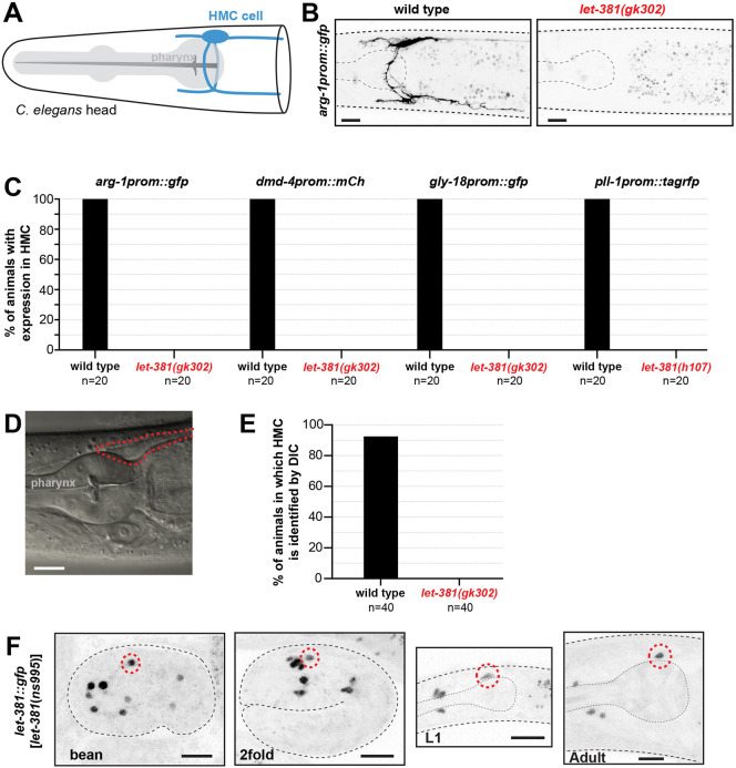
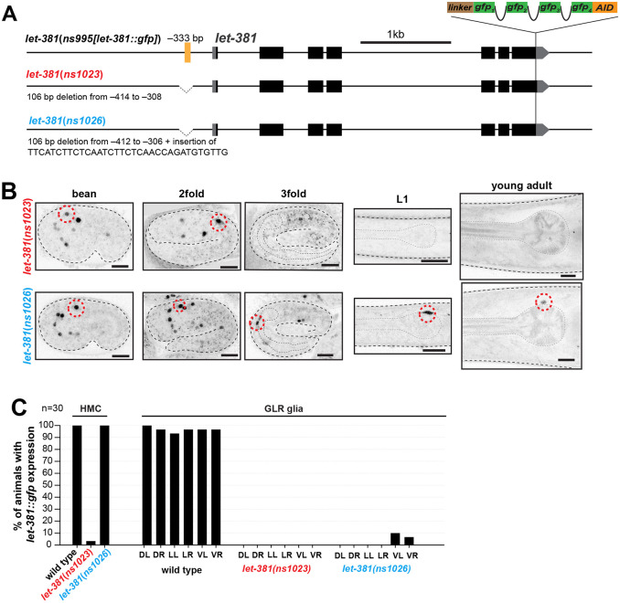
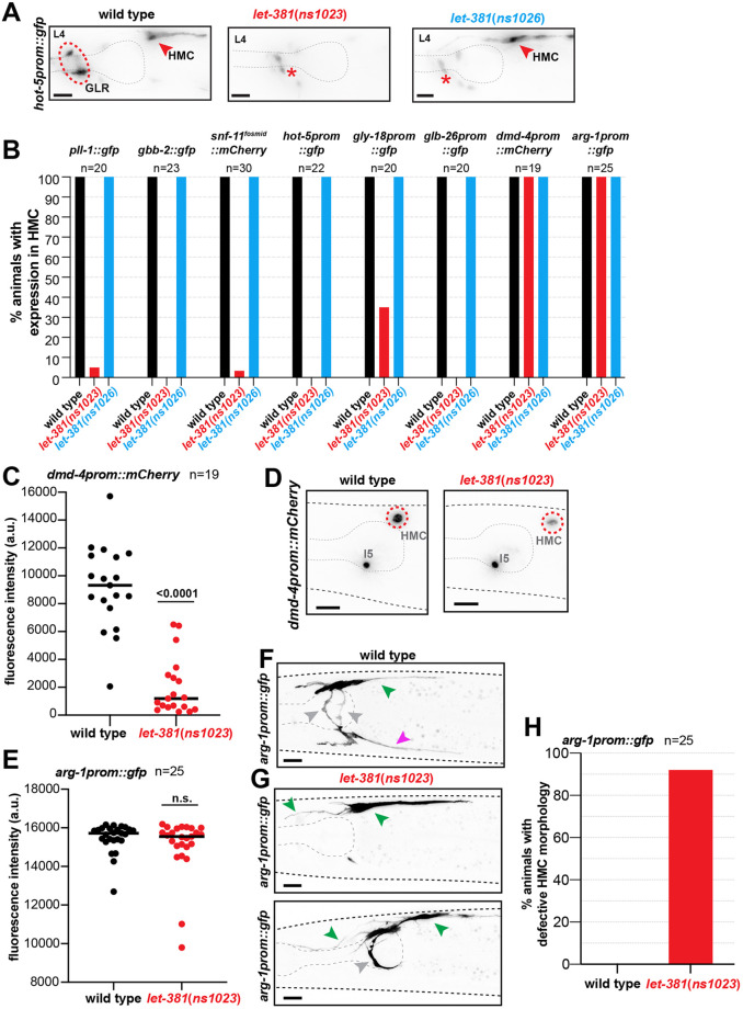
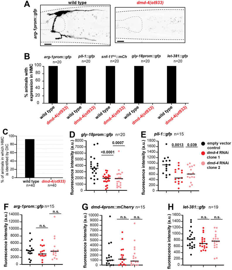
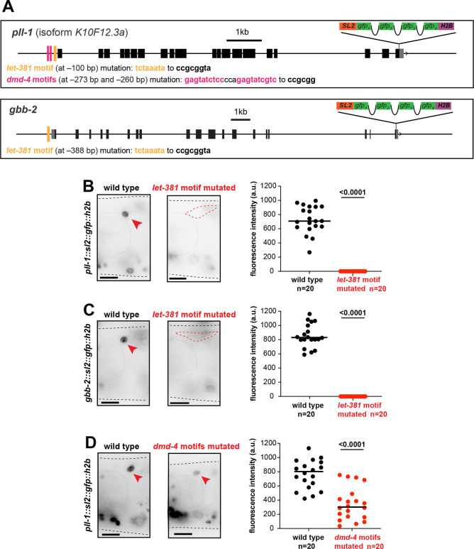
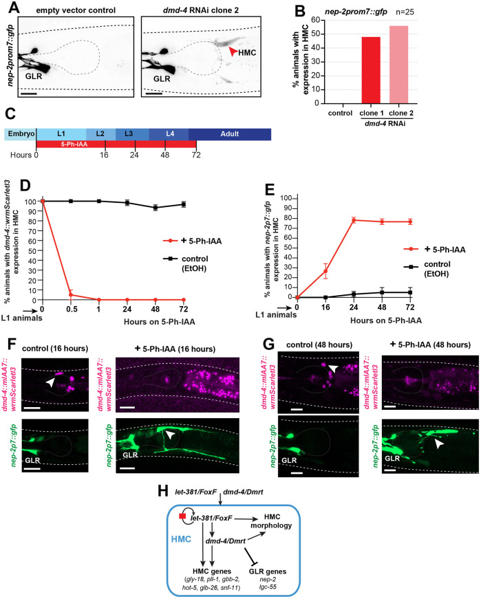
Endothelial cells form the inner layer of blood vessels and play key roles in circulatory system development and function. A variety of endothelial cell types have been described through gene expression and transcriptome studies; nonetheless, the transcriptional programs that specify endothelial cell fate and maintenance are not well understood. To uncover such regulatory programs, we studied the C. elegans head mesodermal cell (HMC), a non-contractile mesodermal cell bearing molecular and functional similarities to vertebrate endothelial cells. Here, we demonstrate that a Forkhead transcription factor, LET-381, is required for HMC fate specification and maintenance of HMC gene expression. DMD-4, a DMRT transcription factor, acts downstream of and in conjunction with LET-381 to mediate these functions. Independently of LET-381, DMD-4 also represses the expression of genes associated with a different, non-HMC, mesodermal fate. Our studies uncover essential roles for FoxF transcriptional regulators in endothelial cell development and suggest that FoxF co-functioning target transcription factors promote specific non-contractile mesodermal fates.

Keywords: Caenorhabditis elegans; dmd-4; let-381; DMRT; Endothelial cell development; FoxF.


Conflict of interest statement

Competing interests The authors declare no competing or financial interests.

Figures

Fig. 1.
Fig. 1.
let-381 is required for HMC specification. (A) Schematic representation of the HMC cell (blue). (B) Fluorescence images of the HMC-specific arg-1prom::gfp reporter in wild type (left) and let-381(gk302) mutant (right). Expression is not observed in the mutant. (C) Percentage of animals with expression in HMC of four different reporters (indicated above the bars) for wild type and let-381 mutants. Expression in the HMC is not detected in the mutants. (D) DIC image of young adult wild-type animal posterior head region (see Fig. 1A). (E) Percentage of young adult animals in which the HMC cell body can be identified by DIC microscopy in wild type and let-381(gk302) mutants. (F) Expression of let-381::gfp in embryonic stages (bean, 2fold), L1 larva and adult animals. n=20 animals for each genotype in C and n=40 for each genotype in E. Anterior is left, dorsal is up. Red dashed lines indicate the HMC cell body in D or HMC nucleus in F. Gray dashed lines outline the animal and the pharynx. Scale bars: 10 μm.
Fig. 2.
Fig. 2.
A let-381 autoregulatory motif is required to maintain LET-381 expression in the HMC. (A) Schematics showing details of let-381 autoregulatory motif deletion mutant alleles. Both alleles remove a sequence containing the let-381 autoregulatory motif (yellow line), located at −333 bp from the ATG, but let-381(ns1026) also has an insertion. (B) Images of let-381::gfp shown for bean, 2-fold and 3-fold embryonic stages, L1 larva and young adults for each genotype. The let-381(ns1023) allele (upper panel row) affects maintenance of let-381::gfp expression in both HMC and GLR glia. let-381(ns1026) (lower panel row) affects maintenance only in GLR glia. (C) Percentage of young adult wild-type, let-381(ns1023) and let-381(ns1026) animals with let-381::gfp expression in HMC and in each of the six GLR glia. let-381::gfp expression is nearly abolished from both the HMC and GLR glia in let-381(ns1023). n=30 animals for each genotype scored in C. Anterior is left, dorsal is up. Red dashed lines indicate the HMC nucleus. Gray dashed lines outline the animal and the pharynx. Scale bars: 10 μm.
Fig. 3.
Fig. 3.
LET-381 is required for maintenance of HMC expression and morphology. (A) hot-5prom::gfp is expressed in both HMC (red arrowhead) and GLR glia (red dashed circle) in wild-type animals. Expression of this reporter is lost in both cell types in let-381(ns1023) animals. By contrast, expression is lost only GLR glia in let-381(ns1026) animals. Red asterisk shows unrelated expression of hot-5prom::gfp in neurons. (B) Percentage of animals with expression in HMC of eight different reporters (indicated above the bars) for wild type, let-381(ns1023) and let-381(ns1026). (C,D) Quantification of fluorescence intensity of dmd-4prom::mCherry reporter in HMC in wild-type and let-381(ns1023) animals (C). dmd-4prom::mCherry expression is significantly reduced in let-381(ns1023). Representative images for each genotype shown in D. (E-G) Quantification of fluorescence intensity of the arg-1prom::gfp reporter in wild-type and ns1023 animals. Fluorescence intensity in the cell body is not affected in let-381(ns1023) (E). Representative images for each genotype shown in F and G. Ventral process (magenta arrowhead) is missing, dorsal process (green arrowheads) is anteriorly mis-extended and lateral processes (gray arrowhead) are missing (top) or mis-extended (bottom) in let-381(ns1023) animals (G). (H) Percentage of animals with HMC morphology defects in wild type and let-381(ns1023) mutants, as assessed with the arg-1prom::gfp reporter. Number of animals (n) scored for each genotype for each reporter is shown under or next to reporter transgene name in B, C, E and G. Black lines on dot-plots indicate mean (C,E). Unpaired t-test was used for statistical analyses in C and E. n.s., not significant. a.u., arbitrary units. Anterior is left, dorsal is up. Gray dashed lines outline the animal and the pharynx. Scale bars: 10 μm.
Fig. 4.
Fig. 4.
DMD-4 is required for HMC fate specification and HMC gene expression. (A) HMC-specific arg-1prom::gfp reporter expression in wild-type (left) and dmd-4(ot933) animals (right). Expression is not observed in dmd-4(ot933). (B) Percentage of animals with expression in HMC of five different reporters (indicated above the bars) for wild type and dmd-4(ot933). Expression in the HMC is not detected in the ot933 mutants for all five reporters. (C) Percentage of young adult animals in which the HMC cell body can be identified by DIC microscopy in wild type and dmd-4(ot933) mutants. (D-H) Quantification of fluorescence intensity for gly-18prom::gfp (D), pll-1::gfp (E), arg-1prom::gfp (F), dmd-4prom::mCherry (G) and let-381::gfp (H) reporters in control and dmd-4 RNAi animals. Number of animals (n) scored for each genotype for each reporter is shown under or next to reporter transgene name in B, D-I and under the genotype for C. Black lines on dot-plots indicate mean (D-H). Unpaired t-test was used for statistical analysis in D-H. n.s., not significant. a.u., arbitrary units. Anterior is left, dorsal is up. Gray dashed lines outline the animal and the pharynx. Scale bars: 10 μm.
Fig. 5.
Fig. 5.
let-381 and dmd-4 motifs are required for endogenous gene expression in the HMC. (A) Schematics of gene models, endogenous gfp-based tags and let-381 motifs (yellow bars) and dmd-4 motifs (magenta bars) for the genes pll-1 and gbb-2. Distance from ATG and nucleotide changes for each motif mutation is shown below the gene models. (B,C) Expression of endogenously gfp-tagged pll-1 (B) and gbb-2 (C) in wild-type and let-381 motif-mutated animals. (D) Expression of endogenously gfp-tagged pll-1 in wild-type and dmd-4 motif-mutated animals. For panels B-D, animal images are on the left, quantifications are shown in the dot-plots on the right. Red arrowhead indicates HMC; red dashed circle outlines HMC cell body in cases where HMC expression is not detected. Black lines on dot-plots indicate mean. Unpaired t-test used for statistical analysis. a.u., arbitrary units. Anterior is left, dorsal is up. Gray dashed lines outline the animal and the pharynx. Scale bars: 10 μm.
Fig. 6.
Fig. 6.
DMD-4 is required for repression of GLR glia genes in the HMC. (A) Representative images showing expression of nep-2prom7::gfp in control and dmd-4(RNAi) animals. Red arrowhead indicates HMC. (B) Percentage of animals with ectopic HMC expression of the GLR-specific reporter nep-2prom7::gfp upon dmd-4 RNAi. n=25 animals for each condition. (C) Timeline of DMD-4 auxin inducible degradation for L1 stage animals (L1 stage DMD-4 AID), showing time points relevant for D and E. (D) Quantification of DMD-4 degradation, based on wrmScarletI3 expression in HMC, at different time points for L1 stage DMD-4 AID. (E) Percentage of animals with ectopic HMC expression of the GLR-specific reporter nep-2prom7::gfp upon L1 stage DMD-4 AID at different time points. For D and E, three replicate experiments were performed, with n=20 animals per replicate for each condition. Error bars show standard deviation between replicates. (F,G) Representative images showing dmd-4::mIAA7::wrmScarletI3::mIAA7 (magenta; top) and nep-2p7::gfp (green; bottom) for control and 5-Ph-IAA-treated animals at different times on 5-Ph-IAA. White arrowheads indicate HMC. HMCs with ectopic nep-2p7::gfp have wild-type appearance at 16 h on 5-Ph-IAA, but display defects at 48 h on 5-Ph-IAA. (H) Schematic representation summarizing the regulatory network for HMC development identified in this study. Anterior is left and dorsal is up for all images. Gray dashed lines outline the animal and the pharynx. Scale bars: 10 μm.

Similar articles

References

    1. Allan, D. W. and Thor, S. (2015). Transcriptional selectors, masters, and combinatorial codes: regulatory principles of neural subtype specification. Wiley Interdiscip. Rev. Dev. Biol. 4, 505-528. 10.1002/wdev.191 - DOI - PMC - PubMed
    1. Ambros, V. (2011). MicroRNAs and developmental timing. Curr. Opin. Genet. Dev. 21, 511-517. 10.1016/j.gde.2011.04.003 - DOI - PMC - PubMed
    1. Amin, N. M., Shi, H. and Liu, J. (2010). The FoxF/FoxC factor LET-381 directly regulates both cell fate specification and cell differentiation in C. elegans mesoderm development. Development 137, 1451-1460. 10.1242/dev.048496 - DOI - PMC - PubMed
    1. Arendt, D., Musser, J. M., Baker, C. V. H., Bergman, A., Cepko, C., Erwin, D. H., Pavlicev, M., Schlosser, G., Widder, S., Laubichler, M. D.et al. (2016). The origin and evolution of cell types. Nat. Rev. Genet. 17, 744-757. 10.1038/nrg.2016.127 - DOI - PubMed
    1. Astorga, J. and Carlsson, P. (2007). Hedgehog induction of murine vasculogenesis is mediated by Foxf1 and Bmp4. Development 134, 3753-3761. 10.1242/dev.004432 - DOI - PubMed

MeSH terms

Substances

LinkOut - more resources