

Lattice Anchoring Stabilizes α-FAPbI3 Perovskite for High-Performance X-Ray Detectors
- PMID: 40728710
- PMCID: PMC12307842
- DOI: 10.1007/s40820-025-01856-4
Lattice Anchoring Stabilizes α-FAPbI3 Perovskite for High-Performance X-Ray Detectors
Abstract
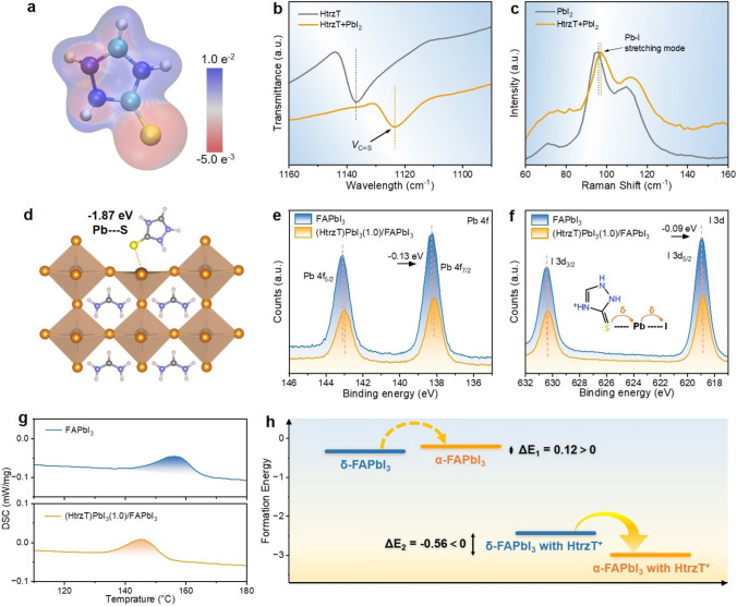
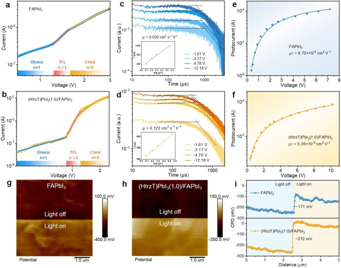
Formamidinium lead iodide (FAPbI3) perovskite exhibits an impressive X-ray absorption coefficient and a large carrier mobility-lifetime product (µτ), making it as a highly promising candidate for X-ray detection application. However, the presence of larger FA+ cation induces to an expansion of the Pb-I octahedral framework, which unfortunately affects both the stability and charge carrier mobility of the corresponding devices. To address this challenge, we develop a novel low-dimensional (HtrzT)PbI3 perovskite featuring a conjugated organic cation (1H-1,2,4-Triazole-3-thiol, HtrzT+) which matches well with the α-FAPbI3 lattices in two-dimensional plane. Benefiting from the matched lattice between (HtrzT)PbI3 and α-FAPbI3, the anchored lattice enhances the Pb-I bond strength and effectively mitigates the inherent tensile strain of the α-FAPbI3 crystal lattice. The X-ray detector based on (HtrzT)PbI3(1.0)/FAPbI3 device achieves a remarkable sensitivity up to 1.83 × 10⁵ μC Gyair-1 cm-2, along with a low detection limit of 27.6 nGyair s-1, attributed to the release of residual stress, and the enhancement in carrier mobility-lifetime product. Furthermore, the detector exhibits outstanding stability under X-ray irradiation with tolerating doses equivalent to nearly 1.17 × 10⁶ chest imaging doses.
Keywords: Conjugated organic cation; Lattice anchoring; Phase stability; X-ray detectors; α-FAPbI3 perovskite.
© 2025. The Author(s).
Conflict of interest statement
Declarations. Conflict of interest: The authors declare no interest conflict. They have no known competing financial interests or personal relationships that could have appeared to influence the work reported in this paper.
Figures


Similar articles
-
The orientation design of high-polarity ligand dipole CF3-PEA for enhancing the surface stability and optoelectronic properties of the FAPbI3 perovskite.Phys Chem Chem Phys. 2025 Jun 25;27(25):13653-13661. doi: 10.1039/d5cp00612k. Phys Chem Chem Phys. 2025. PMID: 40521943
-
Stable and Ultrasensitive X-Ray Detectors Based on Oriented Single-Crystal Perovskite Rods.Adv Mater. 2025 Jul;37(27):e2500101. doi: 10.1002/adma.202500101. Epub 2025 Apr 23. Adv Mater. 2025. PMID: 40269573
-
Intercalation Electrode and Grain Reconstruction Induce Significant Sensitivity Enhancement for Perovskite X-ray Detectors.ACS Appl Mater Interfaces. 2024 Oct 16;16(41):55705-55714. doi: 10.1021/acsami.4c10343. Epub 2024 Oct 4. ACS Appl Mater Interfaces. 2024. PMID: 39364808
-
Impact of residual disease as a prognostic factor for survival in women with advanced epithelial ovarian cancer after primary surgery.Cochrane Database Syst Rev. 2022 Sep 26;9(9):CD015048. doi: 10.1002/14651858.CD015048.pub2. Cochrane Database Syst Rev. 2022. PMID: 36161421 Free PMC article.
-
123I-MIBG scintigraphy and 18F-FDG-PET imaging for diagnosing neuroblastoma.Cochrane Database Syst Rev. 2015 Sep 29;2015(9):CD009263. doi: 10.1002/14651858.CD009263.pub2. Cochrane Database Syst Rev. 2015. PMID: 26417712 Free PMC article.
References
-
- W. Zhao, W.G. Ji, A. Debrie, J.A. Rowlands, Imaging performance of amorphous selenium based flat-panel detectors for digital mammography: characterization of a small area prototype detector. Med. Phys. 30(2), 254–263 (2003). 10.1118/1.1538233 - PubMed
-
- T. Takahashi, S. Watanabe, Recent progress in CdTe and CdZnTe detectors. IEEE Trans. Nucl. Sci. 48(4), 950–959 (2001). 10.1109/23.958705
-
- Q. Guan, S. You, Z.-K. Zhu, R. Li, H. Ye et al., Three-dimensional polar perovskites for highly sensitive self-driven X-ray detection. Angew. Chem. Int. Ed. 63(11), e202320180 (2024). 10.1002/anie.202320180 - PubMed
LinkOut - more resources
Full Text Sources