

Effect of platelet-rich fibrin on microperfusion during early socket healing: a randomized controlled clinical trial
- PMID: 40728781
- PMCID: PMC12307558
- DOI: 10.1007/s00784-025-06470-7
Effect of platelet-rich fibrin on microperfusion during early socket healing: a randomized controlled clinical trial
Abstract
Objectives: This study aimed to evaluate and compare the early healing of fresh alveolar sockets treated with or without platelet-rich fibrin (PRF) using laser Doppler flowmetry and tissue spectrophotometry (LDF-TS). The primary outcome was gingival perfusion; secondary outcomes included clinical wound healing (based on the Landry Wound Healing Index) and patient-reported postoperative pain.
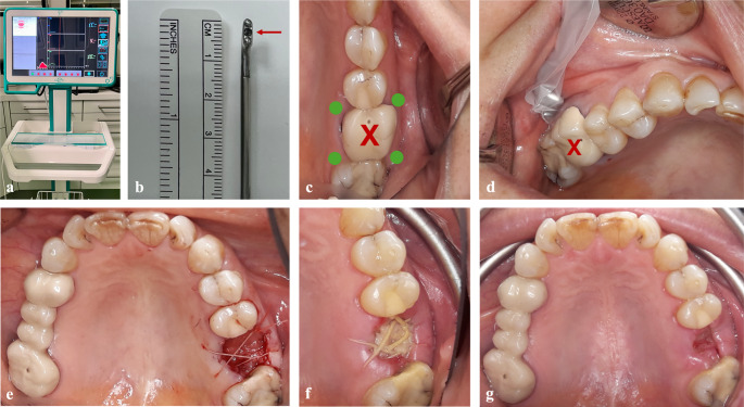
Materials and methods: Sixty-two patients requiring single tooth extraction were randomized into two groups. In the PRF group, an advanced PRF (A-PRF+) plug was placed in the socket before suturing; in the control group, only suturing was performed. Gingival perfusion was measured at four sites preoperatively and on postoperative days 3 and 10 using LDF-TS. Patients rated pain, and wound healing was clinically assessed. Twelve patients were lost to follow-up, leaving 50 for analysis.
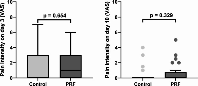
Results: No significant differences were found between the PRF and control group regarding pain (day 3: p = 0.654; day 10: p = 0.329) or wound healing (day 3: p = 0.178; day 10: p = 0.595). Perfusion parameters also showed no significant group differences between baseline and day 10: oxygen saturation (SO₂: p = 0.884), relative hemoglobin (rHb: p = 0.387), and blood flow (p = 0.072).
Conclusions: Gingival perfusion showed no significant group differences over 10 days. PRF did not significantly reduce pain or improve wound healing.
Clinical relevance: PRF does not appear to significantly enhance healing, pain reduction, or perfusion in simple extractions. Future studies should use split-mouth designs and focus on more complex surgeries to better evaluate PRF's effects.
Trial registration: All procedures performed in this study involving human participants were in accordance with the ethical standards of the institutional and/or national research committee and with the 1964 Helsinki Declaration and its later amendments or comparable ethical standards. The study was performed according to the Consolidated Standards of Registered Trial (CONSORT) guidelines. The study was approved by the institutional Clinical Research Ethics Committee (Decision Number 23-105) and by the German Clinical Trials Register (File Number DRKS00032344, registered on October 11, 2023).
Keywords: Laser-Doppler flowmetry; Platelet-rich fibrin; Ridge preservation; Tooth extraction; Wound healing.
© 2025. The Author(s).
Conflict of interest statement
Declarations. Consent for publication: The patients signed informed consent regarding publication of their data and photographs. Competing interests: The authors declare no competing interests. Ethical approval: All procedures performed in this study involving human participants were in accordance with the ethical standards of the institutional and/or national research committee and with the 1964 Helsinki Declaration and its later amendments or comparable ethical standards. The study was performed according to the Consolidated Standards of Registered Trial (CONSORT) guidelines. The study was approved by the institutional Clinical Research Ethics Committee (Decision Number 23–105) and by the German Clinical Trials Register (File Number DRKS00032344, registered on October 11, 2023). Consent to participate: Informed consent was obtained from all individual participants included in the study.
Figures


References
-
- Cardaropoli G, Araújo M, Lindhe J (2003) Dynamics of bone tissue formation in tooth extraction sites. An experimental study in dogs. J Clin Periodontol 30(9):809–818. 10.1034/j.1600-051x.2003.00366.x - PubMed
-
- Araújo MG, Dias DR, Matarazzo F (2023) Anatomical characteristics of the alveolar process and basal bone that have an effect on socket healing. Periodontol 2000 93(1):277–288. 10.1111/prd.12506 - PubMed
-
- Hämmerle CH, Araújo MG, Simion M (2012) Evidence-based knowledge on the biology and treatment of extraction sockets. Clin Oral Implants Res 23 Suppl 5:80–82. 10.1111/j.1600-0501.2011.02370.x - PubMed
-
- Araújo MG, Silva CO, Misawa M, Sukekava F (2015) Alveolar socket healing: what can we learn? Periodontol 2000. Periodontol 2000 68(1):122–134. 10.1111/prd.12082 - PubMed
-
- Iorio-Siciliano V, Ramaglia L, Blasi A, Bucci P, Nuzzolo P, Riccitiello F, Nicolò M (2020) Dimensional changes following alveolar ridge preservation in the posterior area using bovine-derived xenografts and collagen membrane compared to spontaneous healing: a 6-month randomized controlled clinical trial. Clin Oral Investig 24(2):1013–1023. 10.1007/s00784-019-02979-w - PubMed
Publication types
MeSH terms
LinkOut - more resources
Full Text Sources
Miscellaneous
