. 2025 Jul 4;47(7):520.
doi: 10.3390/cimb47070520.

Restoration of Autophagy and Apoptosis in Myelodysplastic Syndromes: The Effect of Azacitidine in Disease Pathogenesis

Affiliations

Restoration of Autophagy and Apoptosis in Myelodysplastic Syndromes: The Effect of Azacitidine in Disease Pathogenesis

Georgia Tsekoura et al. Curr Issues Mol Biol. .

Abstract

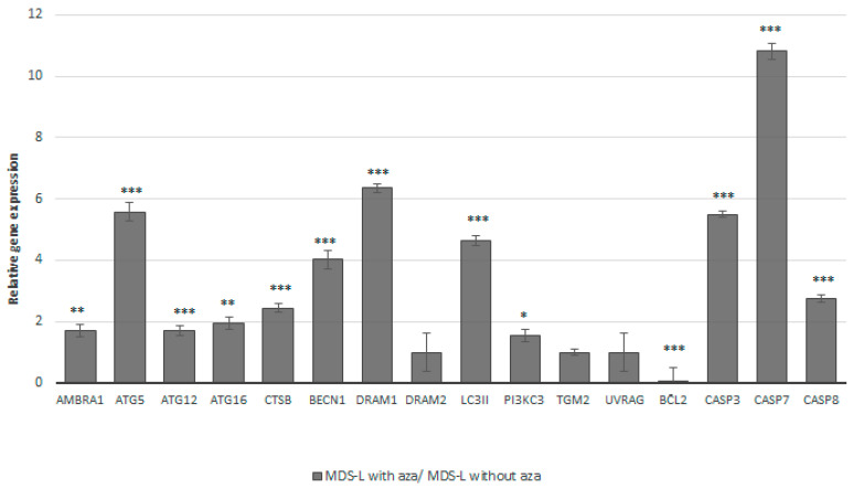
Myelodysplastic syndromes (MDSs) comprise a diverse group of clonal hematopoietic stem cell disorders characterized by ineffective hematopoiesis, cytopenia in the peripheral blood, and an increased risk of transformation into acute myeloid leukemia (AML). Despite extensive research, the mechanisms underlying MDS pathogenesis remain unclear. In the present study, we explored the role of autophagy and apoptosis in the development of MDS and assessed the impact of azacitidine on these processes in vitro. First, we assessed the expression of proteins involved in both autophagic and apoptotic pathways in MDS patients with different prognoses. Furthermore, using the MDS-L cell line as a model, we investigated the in vitro effects of azacitidine treatment on these processes. We report that MDS, irrespective of risk classification, is associated with the dysregulation of autophagy and apoptosis. Notably, azacitidine treatment restored these cellular processes, accompanied by modulation of key signaling phosphoproteins. Overall, these findings provide evidence that impaired autophagy and apoptosis contribute to MDS pathogenesis and that azacitidine helps restore cellular homeostasis by activating both processes. Furthermore, our study highlights the potential therapeutic benefits of targeting these mechanisms and suggests that combining azacitidine with agents that modulate autophagy and apoptosis could enhance the treatment efficacy for MDS patients.

Keywords: apoptosis; autophagy; azacitidine; myelodysplastic syndromes; protein expression profiling.


Conflict of interest statement

Authors Vaia Pliaka and Leonidas Alexopoulos were employed by the company Protavio Ltd. The remaining authors declare that the research was conducted in the absence of any commercial or financial relationships that could be construed as a potential conflict of interest.

Figures

Figure 1
Figure 1
Lower expression levels of proteins implicated in the processes of autophagy and apoptosis in MDS patients compared to those in healthy donors. (A) Western blot experiments in representative samples from the groups of healthy donors (Ctrl1, Ctrl2) and MDS patients of different classifications (HR-MDS: HR1, HR2; LR-MDS: LR1, LR2). The intensity of the bands was quantified using the ImageJ 1.x software, while the expression ratio of each protein was estimated relative to that for the GAPDH protein. (B) Relative expression levels in the group of all MDS patients as well as in each of the MDS risk groups compared to those in the healthy controls. The statistical examination was performed using a one-way ANOVA and Tukey’s post hoc test. Asterisks represent statistically significant differences; one asterisk (*) corresponds to p < 0.05, two asterisks (**) correspond to p < 0.02, and three asterisks (***) correspond to p< 0.001. Error bars correspond to the standard deviation (SD).
Figure 2
Figure 2
Treatment with azacitidine can induce strong cell apoptosis in MDS-L cells even after 24 h of culture. The graph depicts the cell viability curves for MDS-L cells treated with different concentrations of azacitidine (0.5 μΜ, 1 μΜ, 3 μΜ, or 5 μM) after 24, 48, and 72 h. Higher concentrations of azacitidine demonstrated an enhanced cytotoxic effect on the cells, with the degree of cytotoxicity progressively increasing over time, independent of the concentration. Asterisks represent statistically significant differences corresponding to p < 0.05. Error bars correspond to the standard errors (SEs).
Figure 3
Figure 3
Treatment of MDS-L cells with a low concentration of azacitidine may lead to the restoration of autophagy and apoptosis. The expression patterns for a selected panel of 16 genes related to the processes of autophagy and apoptosis in MDS-L cells. Bars represent the relative gene expression levels in the MDS-L cells after treatment with azacitidine compared to those under the baseline status (i.e., without azacitidine treatment). Azacitidine treatment led to upregulation of all of the genes, besides downregulating the anti-autophagic and anti-apoptotic BCL2 gene. The HPRT1 gene was used as a reference. Asterisks represent statistically significant differences; one asterisk (*) corresponds to p < 0.05, two asterisks (**) correspond to p < 0.02, and three asterisks (***) correspond to p< 0.001. Error bars correspond to the standard errors (SEs).
Figure 4
Figure 4
Azacitidine treatment can promote the activation of autophagy by upregulating promoting proteins and downregulating inhibitory proteins. (A) The immunoblot analysis of the expression of a series of proteins implicated in the processes of autophagy and apoptosis in MDS-L cells with and without treatment with azacitidine (MDS-L with aza and MDS-L without aza, respectively). Overall, azacitidine treatment was associated with higher expression levels of the AMPKA, ATG5, BECN1, and LC3II proteins, as well as lower expression levels of BCL2. (B) A bar graph depicting the relative expression levels of all proteins in the treated MDS-L cells compared to those in the baseline (untreated) MDS-L cells. Asterisks represent statistically significant differences; one asterisk (*) corresponds to p < 0.05, two asterisks (**) correspond to p < 0.02, and three asterisks (***) correspond to p < 0.001. Error bars correspond to standard errors (SEs).

References

    1. Garcia-Manero G. Myelodysplastic syndromes: 2023 update on diagnosis, risk-stratification, and management. Am. J. Hematol. 2023;98:1307–1325. doi: 10.1002/ajh.26984. - DOI - PMC - PubMed
    1. McDonald L., McCarthy P., Khan M., Hogan P., Kelleher E.C., Murphy P., Quinn J., Desmond R., McHugh J., Strickland M., et al. Should Myelodysplastic Syndromes in Very Old Patients be More Actively Managed? Clinical Characteristics, Management and Outcomes for Patients 85 Years and Older. Blood. 2018;132((Suppl. 1)):5515. doi: 10.1182/blood-2018-99-113290. - DOI
    1. Watson A.S., Mortensen M., Simon A.K. Autophagy in the pathogenesis of myelodysplastic syndrome and acute myeloid leukemia. Cell Cycle. 2011;10:1719–1725. doi: 10.4161/cc.10.11.15673. - DOI - PMC - PubMed
    1. Parker J.E., Mufti G.J. The Role of Apoptosis in the Pathogenesis of the Myelodysplastic Syndromes. Int. J. Hematol. 2001;73:416–428. doi: 10.1007/BF02994003. - DOI - PubMed
    1. Ames K., Gritsman K. Unraveling the Link between Class 1A PI3-Kinase, Autophagy, and Myelodysplasia. Autophagy. 2024;20:952–954. doi: 10.1080/15548627.2023.2221922. - DOI - PMC - PubMed

LinkOut - more resources