Waldenstrom Macroglobulinemia Recurrence with Bing-Neel Syndrome Presentation
- PMID: 40729145
- PMCID: PMC12225338
- DOI: 10.3390/reports7020034
Waldenstrom Macroglobulinemia Recurrence with Bing-Neel Syndrome Presentation
Abstract
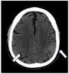
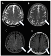
Bing-Neel syndrome (BNS) is a rare condition that may occur in patients with Waldenstrom macroglobulinemia (WM) and is caused by lymphoplasmacytic infiltration into the central nervous system. BNS is an extramedullary manifestation of WM which may present with various neurological signs and symptoms that make the diagnosis difficult to achieve. We present a case of BNS in a 60-year-old patient diagnosed 6 years after recovering from Waldenstrom's macroglobulinemia. We observed the patient for a secondary generalized focal motor seizure. Unenhanced brain CT revealed slight hyperdensity of left parietal subarachnoid spaces. The MRI of the brain and spinal cord showed leptomeningeal enhancement in both parietal lobes. The presence of monoclonal bands (light chain k and IgM) was found in cerebrospinal fluid, leading to the diagnosis of BNS. The patient started treatment with ibrutinib and remains clinically stable during a 1-year follow-up. However, the MRI showed the appearance of a new subcortical left parietal lesion. BNS is an extremely rare presentation of WM that should be recognized and considered early in the presence of unexplained neurological symptoms in patients with a history of WM, even if the patient appears to have recovered.
Keywords: Bing–Neel syndrome; Waldenstrom macroglobulinemia; ibrutinib; leptomeningeal enhancement; monoclonal bands.
Conflict of interest statement
The authors declare that the research was conducted in the absence of any commercial or financial relationships that could be construed as potential conflicts of interest.
Figures


Similar articles
-
Ibrutinib as treatment for Bing-Neel syndrome reclassified as glioblastoma: a case report.J Med Case Rep. 2024 Sep 11;18(1):424. doi: 10.1186/s13256-024-04757-z. J Med Case Rep. 2024. PMID: 39256774 Free PMC article.
-
Bing-Neel syndrome: A rare complication of waldenstrom macroglobulinemia.Indian J Pathol Microbiol. 2025 Apr 1;68(2):405-407. doi: 10.4103/ijpm.ijpm_997_23. Epub 2024 Jun 4. Indian J Pathol Microbiol. 2025. PMID: 38847219
-
Report of Consensus Panel 2 from the 12th International Workshop on the management of Bing-Neel syndrome in patients with Waldenstrom's Macroglobulinemia.Semin Hematol. 2025 Apr;62(2):85-89. doi: 10.1053/j.seminhematol.2025.04.005. Epub 2025 Apr 16. Semin Hematol. 2025. PMID: 40414752
-
Idiopathic (Genetic) Generalized Epilepsy.2024 Feb 12. In: StatPearls [Internet]. Treasure Island (FL): StatPearls Publishing; 2025 Jan–. 2024 Feb 12. In: StatPearls [Internet]. Treasure Island (FL): StatPearls Publishing; 2025 Jan–. PMID: 31536218 Free Books & Documents.
-
¹⁸F-FDG PET/CT: a review of diagnostic and prognostic features in multiple myeloma and related disorders.Clin Exp Med. 2015 Feb;15(1):1-18. doi: 10.1007/s10238-014-0308-3. Epub 2014 Sep 14. Clin Exp Med. 2015. PMID: 25218739
References
-
- Fitsiori A., Fornecker L.M., Simon L., Karentzos A., Galanaud D., Outteryck O., Vermersch P., Pruvo J.-P., Gerardin E., Lebrun-Frenay C., et al. Imaging spectrum of Bing–Neel syndrome: How can a radiologist recognise this rare neurological complication of Waldenström’s macroglobulinemia? Eur. Radiol. 2019;29:102–114. doi: 10.1007/s00330-018-5543-7. - DOI - PubMed
Publication types
LinkOut - more resources
Full Text Sources
