

Intraoral Lipoma on the Palate of an 11-Year-Old Patient: A Case Report
- PMID: 40729232
- PMCID: PMC12199924
- DOI: 10.3390/reports8010019
Intraoral Lipoma on the Palate of an 11-Year-Old Patient: A Case Report
Abstract
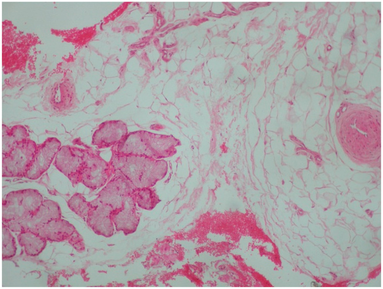
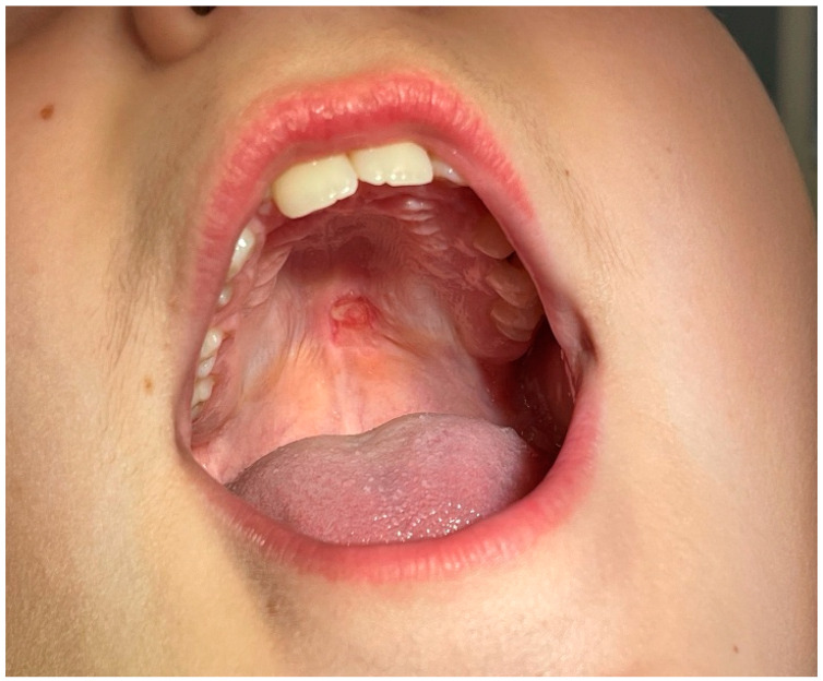
Background and Clinical Significance: Lipomas, benign tumors composed of adipose tissue, are recognized as one of the two most common fat-containing soft tissue tumors, underscoring their relative prevalence among benign tumors in children. Despite their prominence, lipomas rarely occur before 20 years of age, highlighting a discrepancy between their commonality and the age at which they typically manifest. This case report focuses on a 11-year-old patient who noticed the presence of an intraoral mass, which prompted further investigation, ultimately leading to the diagnosis of a lipoma located on the palate. Following our diagnosis, we searched for similar cases; however, the relevant literature was rather limited. There was a case report of a 4-year-old patient who presented with a lipoma on her tongue and a case report of a 6-year-old patient who presented with a lipoma on the buccal mucosa. Case Presentation: The young patient came with his parents to the Department of Oral Medicine and Pathology, School of Dentistry, Aristotle University of Thessaloniki, Greece, and reported the presence of a growth in the middle of the upper jaw. A tumor, of approximately 1 cm diameter, was observed in the middle of the palate, on the border between the hard and soft palate. The surrounding mucosa appeared normal, which is critical in differentiating the tumor from more aggressive pathological entities. It was characterized by a soft and slippery consistency. The patient was referred to a cone beam computed tomography (CBCT) examination to investigate if there was any bone involvement. Based on clinical and radiographical findings, a biopsy was carried out. The tumor was initially excised in its entirety and the base was electrocauterized to avoid placing sutures. The histopathological examination that followed suggested the presence of an intraoral lipoma since lobules of mature adipose tissue in lamina propria and fatty tissue in close proximity to mucinous salivary glands were noticed. Conclusions: The development of lipomas in young patients can be attributed to a multitude of factors that interplay with one another, emphasizing the need for a comprehensive understanding of these growths. Additionally, underlying conditions such as diabetes mellitus, hypercholesterolemia, and obesity also play a crucial role, highlighting the interconnected nature of metabolic disorders and lipoma formation. The surgical approaches for the removal of oral lipomas primarily revolve around complete surgical excision, which is considered the mainstay treatment for these benign tumors.
Keywords: benign; children; head and neck pathology; intraoral; lesion; lipoma; oral; palatal; pediatric; tumor.
Conflict of interest statement
The authors declare no conflicts of interest.
Figures


References
Publication types
LinkOut - more resources
Full Text Sources