

One Family with Cholestasis: The Twisted Road to the Diagnosis of Pfic 3-Three Case Reports
- PMID: 40729246
- PMCID: PMC12199986
- DOI: 10.3390/reports8010033
One Family with Cholestasis: The Twisted Road to the Diagnosis of Pfic 3-Three Case Reports
Abstract
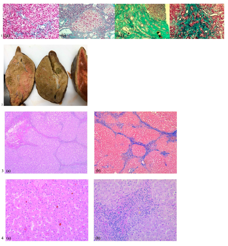
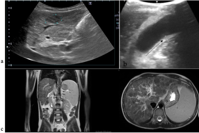
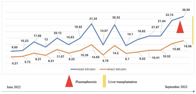
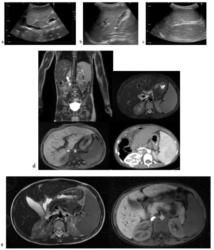
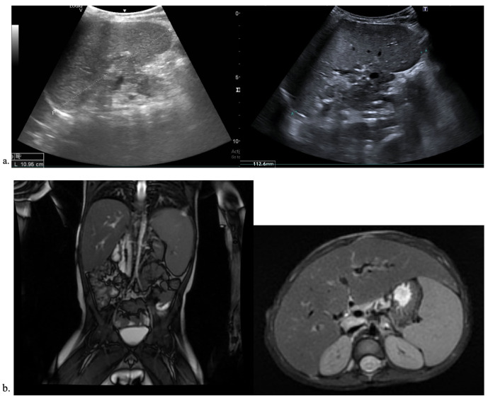
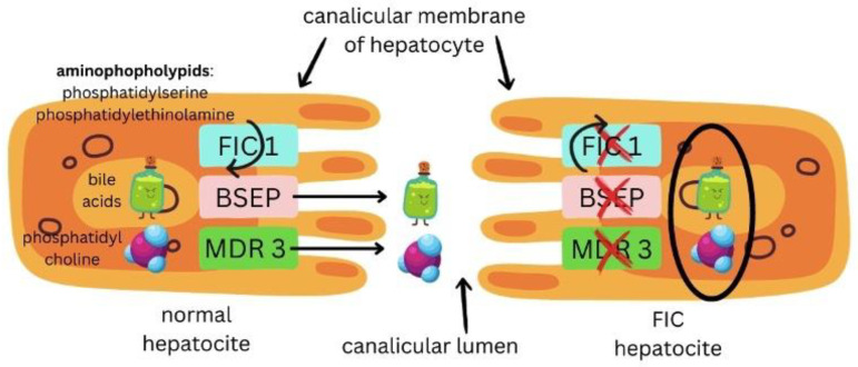
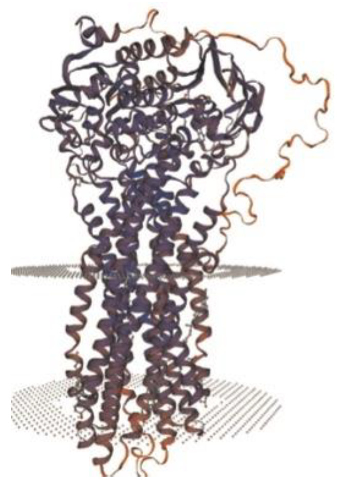
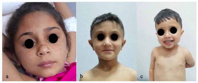
Background and Clinical Significance: Progressive familial intrahepatic cholestasis (PFIC) refers to a heterogeneous group of autosomal recessive disorders consisting of mutations of hepatocyte transporting-system genes involved in bile formation. The exact prevalence remains unknown but is estimated at 1 in 500.000 for PFIC 3, caused by mutations in the ABCB4 gene. We report three cases of PFIC 3 from the patient's sister, brother, and cousin, diagnosed in our Pediatric Department in 2022-2023. Case Presentation: Case 1: A 10-year-old girl was admitted for jaundice and abdominal pain. She was diagnosed with severely advanced hepatic cirrhosis and massive cholestasis. Genetic testing showed ABCB4 homozygous mutation. She rapidly developed fulminant liver failure, and a living donor liver transplant was performed. Case 2: A 6-year-old brother was previously diagnosed with cholestatic hepatitis of unknown cause back in 2018 and presented with similar features (generalized jaundice, severe pruritus with generalized scratching lesions); symptoms had progressively developed from the first year of life. He also exhibited particular facial features (big forehead, twisted ear lobe, straight nose). He received cadaveric liver transplantation. Case 3: Nephew of first two children, a 3-year-5-month-old boy, was admitted for failure to thrive and a one-year history of jaundice, pruritus, and splenomegaly. He was tested positive for homozygous ABCB4 mutation. He is currently under medical treatment with stable liver function. Conclusions: The clinical significance of this particular homozygous variant identified in ABCB4 in our series of cases (c.2534G>T (p.Gly845Val)) was uncertain up to this case report. The present data provide convincing evidence as to the correlation between this mutation and the clinical phenotype of PFIC 3.
Keywords: chronic cholestasis; genetics; jaundice; metabolic liver disease; pruritus; transplantation.
Conflict of interest statement
The authors declare no conflicts of interest.
Figures
Similar articles
-
Epidemiology and burden of progressive familial intrahepatic cholestasis: a systematic review.Orphanet J Rare Dis. 2021 Jun 3;16(1):255. doi: 10.1186/s13023-021-01884-4. Orphanet J Rare Dis. 2021. PMID: 34082807 Free PMC article.
-
Maralixibat in progressive familial intrahepatic cholestasis (MARCH-PFIC): a multicentre, randomised, double-blind, placebo-controlled, phase 3 trial.Lancet Gastroenterol Hepatol. 2024 Jul;9(7):620-631. doi: 10.1016/S2468-1253(24)00080-3. Epub 2024 May 6. Lancet Gastroenterol Hepatol. 2024. PMID: 38723644 Clinical Trial.
-
Lipolysis-Stimulated Lipoprotein Receptor Gene Variants as a Cause of Progressive Familial Intrahepatic Cholestasis: A Case Report.Hepatol Res. 2025 Jul 18. doi: 10.1111/hepr.70003. Online ahead of print. Hepatol Res. 2025. PMID: 40679108
-
Severe Relapsing Hailey-Hailey Disease Displaying a Durable Complete Response to Hydroxyurea.Acta Dermatovenerol Croat. 2024 Nov;32(3):168-169. Acta Dermatovenerol Croat. 2024. PMID: 40654217
-
Signs and symptoms to determine if a patient presenting in primary care or hospital outpatient settings has COVID-19.Cochrane Database Syst Rev. 2022 May 20;5(5):CD013665. doi: 10.1002/14651858.CD013665.pub3. Cochrane Database Syst Rev. 2022. PMID: 35593186 Free PMC article.
References
-
- Bai J., Li L., Liu H., Liu S., Bai L., Ning H., Song W., Zou H., Wang X., Chen Y., et al. A novel compound heterozygous mutation in ABCB4 gene in a pedigree with progressive familial intrahepatic cholestasis 3: A case report. Ann. Transl. Med. 2021;9:426. doi: 10.21037/atm-20-3747. - DOI - PMC - PubMed
Publication types
LinkOut - more resources
Full Text Sources