. 2025 Aug 19;12(8):ENEURO.0252-25.2025.
doi: 10.1523/ENEURO.0252-25.2025. Print 2025 Aug.

Experience-Dependent Intrinsic Plasticity in Layer IV of Barrel Cortex at Whisking Onset

Affiliations

Experience-Dependent Intrinsic Plasticity in Layer IV of Barrel Cortex at Whisking Onset

Molly C Shallow et al. eNeuro. .

Abstract

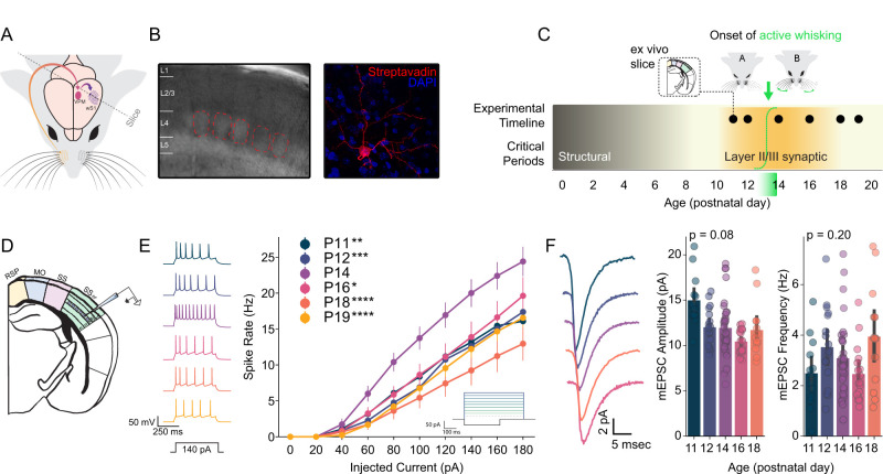
The development of motor control over sensory organs is a critical milestone, enabling active exploration and shaping of the sensory environment. Whether the onset of sensory organ motor control directly influences the development of corresponding sensory cortices remains unknown. Here, we confirm and exploit the late onset of whisking behavior in mice to address this question in the somatosensory system. Using ex vivo electrophysiology, we describe a transient increase in the intrinsic excitability of excitatory neurons in layer IV of the barrel cortex, which processes whisker input, immediately following the onset of active whisking on postnatal days 13 and 14. This increase in neuronal gain is specific to layer IV, independent of changes in synaptic strength, and requires prior sensory experience. Further, these effects are not expressed in inhibitory interneurons in barrel cortex. The transient increase in excitability is not evident in layer II/III of barrel cortex or in the visual cortex upon eye opening, suggesting a unique interaction between the development of active sensing and the thalamocortical input layer in the somatosensory isocortex. Predictive modeling indicates that, immediately following the onset of active whisking, changes in active membrane conductances alone can reliably distinguish neurons in control but not whisker-deprived hemispheres. Our findings demonstrate an experience-dependent, lamina-specific refinement of neuronal excitability tightly linked to the emergence of active whisking. This transient increase in the gain of the thalamic input layer coincides with a critical period for synaptic plasticity in downstream layers, suggesting a role in cortical maturation and sensory processing.

Keywords: barrel cortex; behavior; development; excitability, plasticity.


Conflict of interest statement

The authors declare no competing financial interests.

Figures

Figure 1.
Figure 1.
Developmental onset of active whisking behavior in mice. A, Representative images illustrating three categories of whisking behaviors. Red lines indicate whisker position, with blue arrows showing movement patterns. At P7, mice exhibit non-whisking behavior characterized by largely immobile whiskers. By P10, twitching behavior emerges, characterized by small, non-rhythmic whisker movements. At P16, mice display active whisking with large-amplitude, rhythmic, bilateral whisker movements. B, Stacked bar graph showing the percentage of animals exhibiting each whisking behavior across postnatal days P7–P16. Sample sizes are indicated for each age group. Non-whisking behavior (light blue) dominates at early ages (P7–P8), transitions to predominantly twitching behavior (medium blue) during intermediate ages (P9–P12), and shifts to active whisking (dark blue) at later ages (P13–P16). Complete transition to active whisking occurs by P15. C, Multinomial logistic regression analysis of the developmental transition. Light blue curve represents the probability of non-whisking behavior, medium blue curve represents twitching, and dark blue curve represents active whisking as a function of age. The binary logistic regression equation for active whisking probability is shown [ log(p/(1p))=32.87+2.54×Age, p = 6.28 × 10−8]. The vertical red dashed line indicates the age at which active whisking probability reaches 50% (P12.93), with pink shading representing the 95% confidence interval. Gray shading indicates the age range where active whisking becomes the predominant behavior.
Figure 2.
Figure 2.
Around the onset of active whisking, spiny stellate neurons in layer 4 barrel cortex exhibit a transient increase in excitability. A, Experimental approach. Ex vivo slices were taken from the barrel field of primary somatosensory cortex (SSBF) from postnatal day 11 (P11) through P19. B, Layer IV barrels were visualized under differential interference contrast (DIC) imaging at 10× and individual excitatory cells were chosen under 40× magnification. After recording, cell morphology and location were visualized using streptavidin stains and confocal microscopy. C, Timeline of experiment. Outline of relevant critical periods for structural and synaptic plasticity as well as the timeline of recordings and onset of active whisking (dashed green sigmoid and fade). D, Location of intracellular patch recordings in SSBF. E, Immediately following the onset of active whisking (Fig. 1), P14 F/I curves reveal a transient increase in the intrinsic excitability of Layer IV excitatory cells evident, right. Example traces from each age, left. F, mEPSC recorded from Layer IV excitatory cells do not show an accompanying synaptic strength change, right. Example mEPSC traces from each age, left. Printed p values are for the main effect of age. For F/I curves N = 26 mice, N = 108 cells. For mEPSCs N = 22 mice, N = 94 cells. *p < 0.05, **p < 0.01, ***p < 0.001, ****p < 0.0001. Extended data are presented in Figure 2-1.
Figure 3.
Figure 3.
Increased excitability at the onset of active whisking is specific to excitatory neurons. A, Experimental approach. To fluorescently label a subset of isocortical inhibitory interneurons, Nkx2.1-Cre mice were crossed with TRAP mice, which express EGFP-tagged ribosomal protein L10a under a Cre-dependent promoter. (Top) In this context, Nkx2.1 positive neurons in Layer IV barrels were recorded on postnatal day 12 (p12) through p16. (Bottom) Subsequent staining for SST protein allowed for the separation of three cell types in our dataset: excitatory, PV (NKX positive, SST negative), and SST (NKX and SST positive). B, Example triple label image demonstrating the separation of the three putative cell types. Streptavidin is loaded in the recording electrode and thus labels cells that were patched (intensity reflects the duration of patch). Nkx2.1 labels PV and SST interneurons in the isocortex. SST label is used to define SST interneurons. Merge demonstrates the molecular logic of putative cell type identification. C, PV and SST subpopulations of Nkx2.1 positive neurons are differentiable based on electrophysiological properties, accommodation and the peak instantaneous firing rate. (Top) Kernel density estimate and scatter of Nkx2.1 positive neurons’ accommodation and max firing, colored by PV/SST classification. (Bottom) Example traces from an excitatory neuron (E) and putative PV interneuron in response to a 160 pA current step. D, F/I curve of putative PV interneurons in Layer IV barrel cortex as a function of age reveals no increase in intrinsic excitability on p14. For F/I curves, N = 15 mice, N = 40 cells. Extended data are presented in Figure 3-1.
Figure 4.
Figure 4.
Plasticity in active conductances underlies increased excitability on P14. A, Illustration of recording from excitatory neurons in Layer IV of the barrel fields of primary somatosensory cortex (SSBF). Legend indicates recorded ages and applies to the BJ. BE, Passive properties of SSBF L4 excitatory neurons as a function of age: (B) resting membrane potential, (C) input resistance (measured with a small hyperpolarizing current step), (D) membrane time constant (tau), and (E) membrane capacitance. F, Dynamic input resistance as a function of age. Note that this is a measure of the change in membrane resistance in response to a small positive current, in this case a 40 pA pulse. The voltage deflection is divided by the current (40 pA)—higher values suggest a less leaky membrane. G, Time to first spike as a function of age. H, Afterhyperpolarization (AHP) amplitude as a function of age. I, Spike threshold as a function of age. J, Spike frequency adaptation as a function of age—this refers to a decrease in firing rate over the course of a square wave. KM, Logistic regression was used to predict animal age as a function of three features sets measured in each neuron: active conductances (dynamic input resistance and time to first spike), passive properties (membrane resistance, membrane potential, capacitance, tau), and spiking features (AHP, spike frequency adaptation, and instantaneous frequency). K, Confusion matrix—correctly classified neurons fall on the diagonal—for active conductances. Note that chance is 16.67% and that P14 is robustly identifiable. L, Same as K but for passive properties. M, Same as K but for spiking features.
Figure 5.
Figure 5.
Increased excitability at P14 is not a general property of sensory cortices. A, Inset: Illustration of recording from excitatory neurons in Layer 2/3 of primary somatosensory cortex. Excitatory F/I curves for ages P12, P14, and P16. At P14, L2/3 neurons exhibit reduced excitability compared to P12. B, Excitatory F/I curve of excitatory L4 neurons in the binocular portion of primary visual cortex (V1b) for ages P12, P13, P14, P16, P19, and P20. At P14, neuronal excitability in V1b is unremarkable. C, Mouse eye opening occurs between P10 and P14. F/I curve showing the data presented in B as a function of whether an animal’s eyes were open or closed. For Layer 2/3 F/I curves, N = 11 mice, N = 47 cells. For V1 F/I curves, N = 19 mice, N = 168 cells.
Figure 6.
Figure 6.
Increase in intrinsic excitability is experience-dependent and is blocked by early whisker deprivation. A, Timeline of whisker deprivation. Mice were unilaterally whisker plucked starting at 10 days postnatal (P10) and plucking continued every other day until the day of recording. Ex vivo slices were taken from the barrel field of primary somatosensory cortex (SSBF) from postnatal day 11 (P11) through P19 (B) F/I curves from Layer IV excitatory cells in the deprived hemisphere do not show the transient increase in intrinsic excitability indicating that this change is experience-dependent, insets show the location of intracellular recordings, left, and current injection traces, right. C, Comparing F/I curves for the intact versus deprived hemisphere at each age shows a one day increase in intrinsic excitability in the deprived hemisphere on P11, top left. These comparisons also show that intrinsic excitability in the deprived hemisphere is lower than in the intact hemisphere, further suggesting this change is experience-dependent. * indicates p < 0.05; two-sample t-test of firing frequency at 100 pA current injection. D, F/I curves from Layer II/III excitatory cells in the deprived hemisphere do not show an increase in excitability on P14. Intrinsic excitability in deprived hemisphere Layer II/III cells is slightly higher than in the control hemisphere, and overall excitability decreases with age. E, Confusion matrix showing the ability of a logistic regression classifier to predict an animal’s age based on active conductances in the deprived hemisphere (active conductances comprise dynamic input resistance and time to first spike; correct classifications fall along the diagonal). Note that chance is 16.67% and that P14 is minimally identifiable. F, Classifier balanced accuracy as a function of feature set (gray: active conductances, teal: passive properties, green: spiking features) and condition (intact: solid, deprived: open). Chance is indicated by the dashed red line. For Layer 4 F/I curves, N = 32 mice, N = 142 cells. For Layer 2/3 F/I curves, N = 12 mice, N = 40 cells. Extended data are presented in Figure 6-1.

Similar articles

References

    1. Adelman JP, Maylie J, Sah P (2012) Small-conductance Ca2+-activated K+channels: form and function. Annu Rev Physiol 74:245–269. 10.1146/annurev-physiol-020911-153336 - DOI - PubMed
    1. An S, Yang J-W, Sun H, Kilb W, Luhmann HJ (2012) Long-term potentiation in the neonatal rat barrel cortex in vivo. J Neurosci 32:9511–9516. 10.1523/jneurosci.1212-12.2012 - DOI - PMC - PubMed
    1. Arakawa H, Erzurumlu RS (2015) Role of whiskers in sensorimotor development of C57BL/6 mice. Behav Brain Res 287:146–155. 10.1016/j.bbr.2015.03.040 - DOI - PMC - PubMed
    1. Ayaz A, Stäuble A, Hamada M, Wulf M-A, Saleem AB, Helmchen F (2019) Layer-specific integration of locomotion and sensory information in mouse barrel cortex. Nat Commun 10:2585. 10.1038/s41467-019-10564-8 - DOI - PMC - PubMed
    1. Bender KJ, Rangel J, Feldman DE (2003) Development of columnar topography in the excitatory layer 4 to layer 2/3 projection in rat barrel cortex. J Neurosci 23:8759–8770. 10.1523/jneurosci.23-25-08759.2003 - DOI - PMC - PubMed

LinkOut - more resources