. 2025 Jul 29;15(1):27665.
doi: 10.1038/s41598-025-12571-w.

Derivation of resident macrophages and construction of tumor microenvironment in Flk-1-knockout chimeric mice produced via blastocyst complementation

Affiliations

Derivation of resident macrophages and construction of tumor microenvironment in Flk-1-knockout chimeric mice produced via blastocyst complementation

Satoko Ishii et al. Sci Rep. .

Abstract

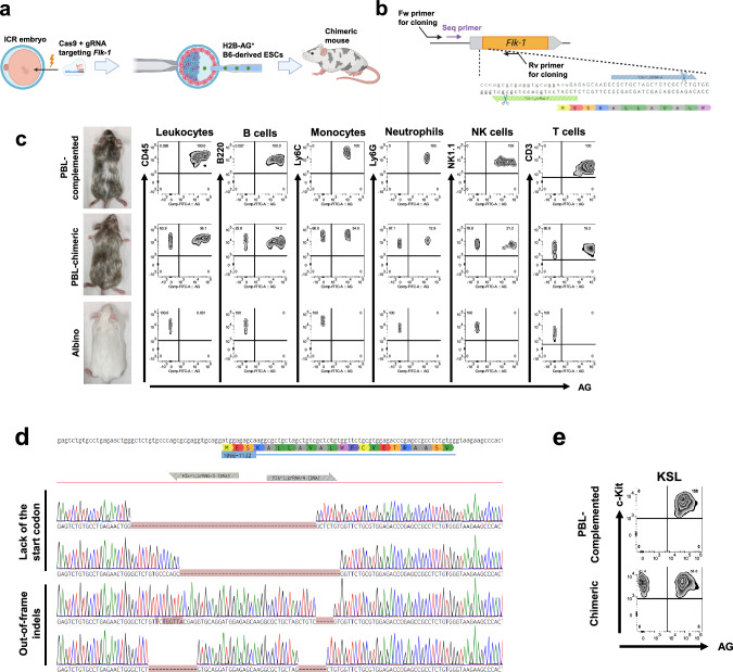
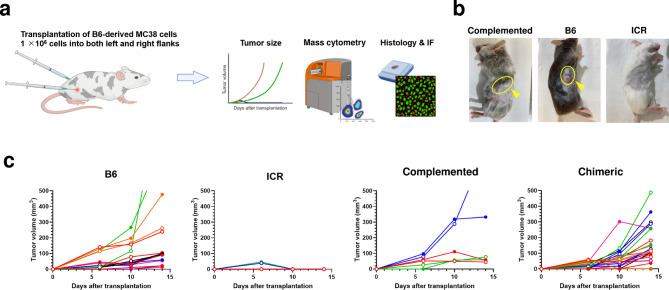
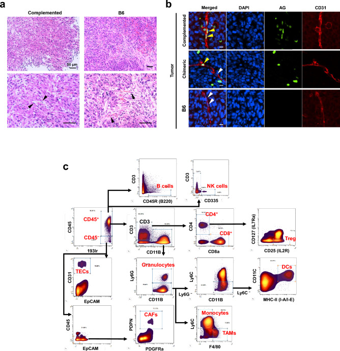
The tumor microenvironment (TME) is deeply involved in cancer progression and treatment resistance. Although humanized mouse models have been developed by transplanting human cells into immunodeficient mice, they fail to fully reconstitute the TME. Blastocyst complementation using Flk-1 (Vegfr2, Kdr) knockout hosts offers a potential solution. However, the generation of interspecies human-mouse chimeras using blastocyst complementation has not yet been successful. As a foundational step, this study aims to demonstrate that donor-derived TME can be constructed using this method in intraspecies chimeric mice. We generated chimeric mice by injecting Azami-Green (AG)-positive C57BL/6 (B6) mouse-derived embryonic stem cells (ESCs) into ICR Flk-1 knockout embryos. We observed that vascular endothelial cells (VECs), hematopoietic cells, and tissue-resident macrophages were derived from the injected AG-positive ESCs. We engrafted B6-derived tumor cells into the chimeras and identified tumor-infiltrating lymphocytes, tumor-associated macrophages, and VECs derived from donor cells. Moreover, tumor-infiltrating CD8+ T cells in these chimeric mice showed cytotoxic activity comparable to that in wild-type mice. We anticipate that this intraspecies chimeric mouse model can serve as a valuable tool for basic research. Furthermore, future humanized tumor models generated via blastocyst complementation have the potential to significantly advance anticancer drug development in the preclinical phase.

Keywords: Blastocyst complementation; Resident macrophages; Tumor microenvironment; Tumor model.


Conflict of interest statement

Declarations. Competing interests: The authors declare no competing interests.

Figures

Fig. 1
Fig. 1
Generation of Flk-1-knockout chimeric mice via blastocyst complementation. (a) Schematic of the method to generate histone H2B (H2B)-Azami-Green (AG)-labeled C57BL/6 (B6) embryonic stem cell (ESC) → Flk-1−/− knockout (KO) chimeric mice. Cas9 protein and guide RNAs (gRNAs) against Flk-1 were electroporated into pronuclei of ICR eggs. The treated eggs were cultured to the blastocyst stage. Flk-1-KO blastocysts were injected with AG-labeled B6 embryonic stem cells (ESCs), grown for 24 h, and then transferred into the uteri of pseudopregnant ICR female mice. (b) The target site of CRISPR-Cas9 to knockout Flk-1 and the positions of primers used for cloning (forward (Fw) and reverse (Rv)) and Sanger sequencing (Seq). (c) Flow cytometry analysis of peripheral blood leukocytes (PBLs) in PBL-complemented (almost all PBLs were AG-positive), PBL-chimeric (PBLs were composed of AG-positive and AG-negative cells), and albino mice from the same littermate. (d) Representative examples of Sanger sequencing results of four chimeric mice which had almost 100% AG-positive PBLs. The genomic and amino acid sequences of Flk-1 around the start codon are shown at the top. Below that, the sequences of four Flk-1 KO host chimeras are shown. The top two chimeras lacked the start codon, and the bottom two had out-of-frame indels. (e) Flow cytometry analysis of the c-Kit+ Sca1+ Lineage (KSL) fraction in PBL-complemented and PBL-chimeric mice.
Fig. 2
Fig. 2
Analysis of vascular endothelial cells (VECs) in H2B-AG+ B6 ESC → Flk-1 KO chimeric mice. Immunohistological analysis of VECs in the kidney (a), brain (c), and pancreas (d) of PBL-complemented and PBL-chimeric mice. The yellow and white arrowheads indicate donor-derived (H2B-AG+ CD31+) and host-derived VECs (H2B-AG CD31+), respectively. Scale bars, 10 µm. (b) Flow cytometry analysis of vascular endothelium of the kidneys in PBL-complemented and PBL-chimeric mice.
Fig. 3
Fig. 3
Tissue-resident macrophages in H2B-AG+ B6 ESC → Flk-1 KO mice. (a) Immunohistological analysis of microglia of PBL-complemented and PBL-chimeric mice. The yellow and white arrowheads indicate donor-derived (H2B-AG+ Iba1+) and host-derived microglia (H2B-AG Iba1+), respectively. Scale bars, 50 µm. (b) Flow cytometry analysis of microglia in PBL-complemented and PBL-chimeric mice. (c) Immunohistological analysis of Iba1+ cells in the liver of PBL-complemented and PBL-chimeric mice. The yellow and white arrowheads indicate donor-derived (H2B-AG+ Iba1+) and host-derived (H2B-AG Iba1+) macrophages, respectively. Scale bars, 10 µm. (d) Flow cytometry analysis of resident macrophages of the pancreas in PBL-complemented and PBL-chimeric mice.
Fig. 4
Fig. 4
Tumor cell inoculation into H2B-AG+ B6 ESC → Flk-1 KO chimeric, B6, and ICR mice. (a) Schematic of tumor cell inoculation and analysis of tumors. MC38 cells, a cancer cell line derived from B6 mice, which have the same origin as the donor embryonic stem cells (ESCs), were transplanted into both the flanks, and tumor sizes were measured over time. Tumors were harvested 14 days after the inoculation and cells that constituted the tumor microenvironment were analyzed histologically and using mass cytometry and immunofluorescence (IF). (b) Representative photographs of implanted tumors on a PBL-complemented and B6 (yellow arrowheads and ellipses) mice. No cancer cells were engrafted in ICR mice. (c) Tumor growth curves for PBL-complemented and PBL-chimeric mice, and B6 and ICR mice.
Fig. 5
Fig. 5
Analysis of the tumor microenvironment (TME) in MC38-derived tumors engrafted in H2B-AG+ B6 ESC → Flk-1 KO chimeric and B6 mice. (a) Hematoxylin and eosin staining of tumors in PBL-complemented (left) and B6 (right) mice. Arrowheads indicate vascular endothelial cells (VECs). Top, × 200; bottom, × 400. Scale bar, 50 µm. (b) Immunohistological analysis of VECs in tumors of PBL-complemented, PBL-chimeric, and B6 mice. The yellow and white arrowheads indicate donor-derived (H2B-AG+ CD31+) and host-derived (H2B-AG CD31+) VECs, respectively. Scale bars, 10 µm. (c) Mass cytometry gating scheme used to identify the cell subsets in the TME. Arrows indicate the gating sequence. We used two panels, one of which analyzed all cells other than cancer-associated fibroblasts (CAFs), and the other for all cells other than natural killer (NK) cells. (d) Percentage of AG-positive cells in each cell population in the tumor determined using mass cytometry. The circle represents the value of one tumor. Red, PBL-complemented; Blue, PBL-chimeric; Black, B6. (e) Percentages of granzyme B-positive cells in total and AG+ CD8+ T cells in tumors of B6 mice and PBL-complemented chimeras, respectively. ns, not significant by Mann–Whitney’s U-test. Means ± S.E.M. n = 8 for B6 group; n = 5 for PBL-complemented group.
Fig. 5
Fig. 5
Analysis of the tumor microenvironment (TME) in MC38-derived tumors engrafted in H2B-AG+ B6 ESC → Flk-1 KO chimeric and B6 mice. (a) Hematoxylin and eosin staining of tumors in PBL-complemented (left) and B6 (right) mice. Arrowheads indicate vascular endothelial cells (VECs). Top, × 200; bottom, × 400. Scale bar, 50 µm. (b) Immunohistological analysis of VECs in tumors of PBL-complemented, PBL-chimeric, and B6 mice. The yellow and white arrowheads indicate donor-derived (H2B-AG+ CD31+) and host-derived (H2B-AG CD31+) VECs, respectively. Scale bars, 10 µm. (c) Mass cytometry gating scheme used to identify the cell subsets in the TME. Arrows indicate the gating sequence. We used two panels, one of which analyzed all cells other than cancer-associated fibroblasts (CAFs), and the other for all cells other than natural killer (NK) cells. (d) Percentage of AG-positive cells in each cell population in the tumor determined using mass cytometry. The circle represents the value of one tumor. Red, PBL-complemented; Blue, PBL-chimeric; Black, B6. (e) Percentages of granzyme B-positive cells in total and AG+ CD8+ T cells in tumors of B6 mice and PBL-complemented chimeras, respectively. ns, not significant by Mann–Whitney’s U-test. Means ± S.E.M. n = 8 for B6 group; n = 5 for PBL-complemented group.
Fig. 6
Fig. 6
Schematic of the tumor microenvironment in the current humanized mice and future possibilities. (Left) This model was created by transplanting human hematopoietic stem cells into immunodeficient mice. The humanized mice had human immune cells. However, vascular endothelial cells and some of resident macrophages were derived from mice. (Right) By blastocyst complementation using Flk-1 KO mice as a host, it is possible to humanize not only immune cells but also vascular endothelial cells (tumor endothelial cells) and resident macrophages (tumor-associated macrophages). Therefore, most of cells in the tumor microenvironment are derived from human cells, as suggested in this study. In the case of Flk-1 knockout, however, cancer-associated fibroblasts are presumed to be in a chimeric state. Blue, mouse cells; Red, human cells.

Similar articles

References

    1. Lee, W. S., Yang, H., Chon, H. J. & Kim, C. Combination of anti-angiogenic therapy and immune checkpoint blockade normalizes vascular-immune crosstalk to potentiate cancer immunity. Exp. Mol. Med.52, 1475–1485 (2020). - PMC - PubMed
    1. Petitprez, F., Meylan, M., De Reyniès, A., Sautès-Fridman, C. & Fridman, W. H. The tumor microenvironment in the response to immune checkpoint blockade therapies. Front. Immunol.11, 784 (2020). - PMC - PubMed
    1. Ma, S. et al. The role of tumor microenvironment in resistance to anti-angiogenic therapy. F1000Research7, 326 (2018). - PMC - PubMed
    1. Wege, A. K. Humanized mouse models for the preclinical assessment of cancer immunotherapy. BioDrugs32, 245–266 (2018). - PubMed
    1. Ireson, C. R., Alavijeh, M. S., Palmer, A. M., Fowler, E. R. & Jones, H. J. The role of mouse tumour models in the discovery and development of anticancer drugs. Br. J. Cancer121, 101–108 (2019). - PMC - PubMed

Substances

LinkOut - more resources