. 2025 Jul 29;16(1):6972.
doi: 10.1038/s41467-025-62309-5.

Autocrine interferon poisoning mediates ADAR1-dependent synthetic lethality in BRCA1/2-mutant cancers

Affiliations

Autocrine interferon poisoning mediates ADAR1-dependent synthetic lethality in BRCA1/2-mutant cancers

Roman M Chabanon et al. Nat Commun. .

Abstract

ADAR1 is an RNA editing enzyme which prevents autoimmunity by blocking interferon responses triggered by cytosolic RNA sensors, and is a potential target in immuno-oncology. However, predictive biomarkers for ADAR1 inhibition are lacking. Using multiple in vitro and in vivo systems, we show that BRCA1/2 and ADAR1 are synthetically lethal, and that ADAR1 activity is upregulated in BRCA1/2-mutant cancers. ADAR1 depletion in BRCA1-mutant cells causes an increase in R-loops and consequently, an upregulation of cytosolic nucleic acid sensing pattern recognition receptors (PRR), events which are associated with a tumor cell-autonomous type I interferon and integrated stress response. This ultimately causes autocrine interferon poisoning. Consistent with a key role of R-loops in this process, exogenous RNase H1 expression reverses the synthetic lethality. Pharmacological suppression of cell-autonomous interferon responses or transcriptional silencing of cytosolic nucleic acid sensing PRR are also sufficient to abrogate ADAR1 dependency in BRCA1-mutant cells, in line with autocrine interferon poisoning playing a central part in this synthetic lethality. Our findings provide a preclinical rationale for assessing ADAR1-targeting agents in BRCA1/2-mutant cancers, and introduces a conceptually novel approach to synthetic lethal treatments, which exploits tumor cell-intrinsic cytosolic immunity as a targetable vulnerability of cancer cells.


Conflict of interest statement

Competing interests: R.M.C. makes the following disclosures: is named inventor on a patent describing the use of ADAR1 inhibitors as anti-cancer agents (WO/2024/189433). J.Z. makes the following disclosures: has received research funding and travel subsidies from Menarini. B.P. makes the following disclosures: has received research funding as part of an institutional program from: Astra Zeneca, Daiichi Sankyo, Gilead, Seagen, MSD; receives and/or has received consulting fees from: Astra Zeneca (institutional), Seagen (institutional), Gilead (institutional), Novartis (institutional), Lilly (institutional), MSD (institutional), Pierre Fabre (personal), Daiichi Sankyo (institutional/personal), Olema (institutional); has received travel subsidies from: Astra Zeneca; Pierre Fabre; MSD; Daiichi Sankyo, Pfizer. A.N.J.T. makes the following disclosures: Receipt of grants / research support: AstraZeneca research costs associated with OlympiA, Myriad Genetics research support for TNT Trial, Breast Cancer Now research grant to Institution, CRUK research grant to Institution. Receipt of honoraria or consultation fees: AstraZeneca, Inbiomotion, Gilead, Innovation in Breast Cancer Symposium, SABCS, GBCC, Cancer Panel, Research to Practise, AACR, Aicme, Penn Medicine, PAGE Therapeutics, Ellipses, VHIO, Dana Faber David Livingston Memorial Symposium, Tango Therapuetics, Guardant, CRUK, Neogenomics, Merck. Educational seminar: Guardant. Stock option: InBioMotion. Royalties: Benefits from ICR’s Inventors Scheme associated with patents for use of several PARP inhibitors in DNA repair-deficient cancers held by AstraZeneca with royalty payments to A.N.J.T.’s personal account and research accounts at the Institute of Cancer Research, and to the Institute of Cancer Research. Institutional financial interests: Royalties as above from AstraZeneca. Research costs from AstraZeneca, Myriad Genetics. Non-financial interests: Leadership/Guideline Advisor roles: Director of Breast Cancer Now Research Centre—ICR/KCL, St Gallen Early Breast Cancer Guidelines Panel, ESMO Early Breast Cancer Guidelines Committee, ESMO Scientific Committee, Chair—CRUK Clinical Research Committee, BCRF SAB member, Strategy Committee UK NCRI, Trustee The Cridlan Ross Smith Charitable Trust. C.J.L. makes the following disclosures: receives and/or has received research funding from: AstraZeneca, Merck KGaA, Artios, Neophore; has received consultancy, SAB membership or honoraria payments from: FoRx, Syncona, Sun Pharma, Gerson Lehrman Group, Merck KGaA, Vertex, AstraZeneca, Tango Therapeutics, 3rd Rock, Ono Pharma, Artios, Abingworth, Tesselate, Dark Blue Therapeutics, Pontifax, Astex, Neophore, Glaxo Smith Kline, Dawn Bioventures, Blacksmith Medicines, ForEx; has stock in: Tango, Ovibio, Hysplex, Tesselate, Ariceum. C.J.L. is also named inventor on patents describing the use of DNA repair inhibitors and stands to gain from their development and use as part of the ICR “Rewards to Inventors” scheme and also reports benefits from this scheme associated with patents for PARP inhibitors paid into C.J.L.’s personal accounts and research accounts at the Institute of Cancer Research. C.J.L. is also named inventor on a patent describing the use of ADAR1 inhibitors as anti-cancer agents (WO 2024/189433 A1). S.P.V. makes the following disclosures: has received research funding from Hoffman La Roche and AstraZeneca for unrelated research projects. As part of the Drug Development Department (DITEP), S.P.V. is principal investigator or sub-investigator of clinical trials from Abbvie, Agios Pharmaceuticals, Amgen, Argen-X Bvba, Arno Therapeutics, Astex Pharmaceuticals, Astra Zeneca, Aveo, Bayer Healthcare Ag, Bbb Technologies Bv, Blueprint Medicines, Boehringer Ingelheim, Bristol Myers Squibb, Celgene Corporation, Chugai Pharmaceutical Co., Clovis Oncology, Daiichi Sankyo, Debiopharm S.A., Eisai, Eli Lilly, Exelixis, Forma, Gamamabs, Genentech, Inc., GlaxoSmithKline, H3 Biomedicine, Inc, Hoffmann La Roche Ag, Innate Pharma, Iris Servier, Janssen Cilag, Kyowa Kirin Pharm. Dev., Inc., Loxo Oncology, Lytix Biopharma As, Medimmune, Menarini Ricerche, Merck Sharp & Dohme Chibret, Merrimack Pharmaceuticals, Merus, Millennium Pharmaceuticals, Nanobiotix, Nektar Therapeutics, Novartis Pharma, Octimet Oncology Nv, Oncoethix, Onyx Therapeutics, Orion Pharma, Oryzon Genomics, Pfizer, Pharma Mar, Pierre Fabre, Roche, Sanofi Aventis, Taiho Pharma, Tesaro Inc, and Xencor. S.P.V. has participated to advisory boards for Merck KGaA. S.P.V. is also named inventor on a patent describing the use of ADAR1 inhibitors as anti-cancer agents (WO 2024/189433 A1). All other authors have no conflicts of interest or financial interests to disclose.

Figures
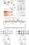
Fig. 1
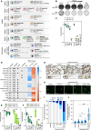
Fig. 1. A functional screen identifies BRCA1ADAR1 synthetic lethality in triple-negative breast cancer.
A Schematic showing experimental design for the generation of SUM149 BRCA1-isogenic cell lines. B, C A focused siRNA screen (B) identifies BRCA1–ADAR1 synthetic lethality in the SUM149 BRCA1-isogenic model (C). Box-and-whiskers indicate median, lower and upper quartiles, and the min to max range; n = 4 biological replicates, two-way ANOVA post hoc Šidák’s test. P value, ****< 0.0001. D, E Clonogenic survival of SUM149 BRCA1-Mut and BRCA1-Rev cells transfected with ADAR1 siRNA (P, Pool; #1; #2). siCTRL, non-targeting, negative control siRNA; siPLK1, PLK1-targeting, positive control siRNA. Violin plots indicate median, lower and upper quartiles; N = 6 values from individual wells, representative of n = 3 biologically-independent experiments, two-way ANOVA post hoc Dunnett’s test. P values, *=0.0196, ****< 0.0001. F, G Clonogenic survival of SUM149 BRCA1-Mut and BRCA1-Rev cells transfected with ADAR1 sgRNA (#1; #2; #3; #4). sgCTRL, non-targeting, negative control sgRNA. Violin plots indicate median, lower and upper quartiles; N = 3 values from individual wells, representative of n = 3 biologically-independent experiments, two-way ANOVA post hoc Dunnett’s test. P values, **=0.0015, ****< 0.0001. Source data are provided as a Source Data file. Elements of panels A and B were provided by Servier Medical Art (https://smart.servier.com/) and BioRender (https://www.biorender.com/), licensed under CC BY 4.0 (https://creativecommons.org/licenses/by/4.0/).
Fig. 2
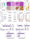
Fig. 2. ADAR1 silencing is synthetically lethal with BRCA1/2 mutations in multiple model systems in vitro and in vivo.
A Schematic describing the isogenic and non-isogenic cell line models used throughout the study. B, C Clonogenic survival of MEF Brca1-wildtype (WT) and Brca1-mutant (Δ11) cells transfected with a concentration range (nM) of Adar1 siRNA. Violin plots indicate median, lower and upper quartiles; N = 6 values from individual wells, representative of n = 3 biologically-independent experiments, two-way ANOVA post hoc Dunnett’s test. P values, *=0.0402, **=0.0017, ****< 0.0001. D Heatmap showing surviving fractions elicited by ADAR1 suppression in models evaluated in Figs. 1, 2 and Supplementary Fig. 1–3 (blue, SF > 0.8; red, SF < 0.8). E, F Cell survival of HEK293T ADAR1-wildtype (WT) and ADAR1-knockout (KO) cells transfected with a concentration range (nM) of BRCA1 (E) or BRCA2 (F) siRNA. Box-and-whiskers indicate median, lower and upper quartiles, and the min to max range; N = 4 values from individual wells, representative of n = 3 biologically-independent experiments, two-way ANOVA post hoc Dunnett’s test. P values, **[ADAR1-WT, siBRCA2 2 nM]=0.001 (F), ***[ADAR1-KO, siBRCA1 2 nM]=0.0009 (E), ***[ADAR1-KO, siBRCA1, 4 nM]=0.0005 (E), ****<0.0001. G–J Representative images and quantifications of morphological phenotypes (G, I) and acridine orange staining (H, J) in zebrafish embryos subjected to morpholino (MO)-mediated knockdown of brca2 and/or adar1. Dead embryos are indicated with an asterisk (G). White arrows indicate acridine orange-positive cells on images taken within a defined region along the anterior-posterior axis (H); scale bar, 500 µm. Percentages of morphological phenotypes (I) were calculated based on N = 149 [no MO], N = 95 [control MO], N = 109 [brca2 MO], N = 97 [adar1 MO] and N = 174 [brca2/adar1 MOs] values from individual embryos, representative of n = 3 biologically-independent clutches. Violin plots indicate median, lower and upper quartiles; N = 31 [no MO], N = 33 [control MO], N = 25 [brca2 MO], N = 23 [adar1 MO] and N = 24 [brca2/adar1 MOs] values from individual embryos, representative of n = 3 biologically-independent experiments, Kruskal-Wallis test post hoc Dunn’s test. P value, ****<0.0001. siCTRL, non-targeting, negative control siRNA; siPLK1, PLK1-targeting, positive control siRNA. Source data are provided as a Source Data file. Elements of panel A were provided by Servier Medical Art (https://smart.servier.com/), licensed under CC BY 4.0 (https://creativecommons.org/licenses/by/4.0/).
Fig. 3
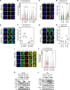
Fig. 3. BRCA1/2 mutations in patients associate with increased tumor cell ADAR1 expression and activity.
A, B Pathological evaluation (A) and representative images (B) of ADAR1p150 cytoplasmic expression according to BRCA1 gene status (BRCA1-wildtype vs. BRCA1-mutant) in a cohort of 63 treatment-naïve triple-negative breast cancer (TNBC) patients. H-score of ADAR1p150 expression (range, 0–300). Violin plots indicate median, lower and upper quartiles; N = 32 [BRCA1-wildtype], N = 31 [BRCA1-mutant] values from individual tumor samples, two-tailed Mann-Whitney U test. P value, *=0.0359. Hematoxylin and eosin (H&E) and ADAR1p150 staining (magnification, ×20) are shown. Scale bars, 50 μm. C Percentage of TILs in TNBC tumors from the cohort described in A, according to cytoplasmic ADAR1p150 expression (based on A; ADAR1p150-low, lower quartile of H-score; ADAR1p150-high, upper quartile of H-score). Violin plots indicate median, lower and upper quartiles; N = 17 [BRCA1-wildtype], N = 15 [BRCA1-mutant] values from individual tumor samples, two-tailed Mann-Whitney U test. P value, **=0.0043. D, E Schematics illustrating the conceptual approach (D) and bioinformatic pipelines (E) used to evaluate A-to-I RNA editing levels from RNA-Seq data. F, G. A-to-I RNA editing levels displayed as RNA editing index (F) or number of RNA editing sites (G) in SUM149 BRCA1-Mut and BRCA1-Rev cells transfected with ADAR1 siRNA (P, Pool; #1). siCTRL, non-targeting, negative control siRNA. Bar plots indicate mean ± SD; n = 3 biological replicates, two-way ANOVA post hoc Tukey’s test. P values, ****< 0.0001. H Schematic of clinical history of the BRCA2-mutant and -revertant patient-derived xenografts (PDXs) MR-0009 and MR-0191; arrows indicate times of tumor biopsies for PDX establishment. Duration of each treatment delivered after diagnosis is indicated in months. Details of the corresponding BRCA2 mutations (germline vs. reversion) are presented to the left. I, J A-to-I RNA editing levels displayed as RNA editing index (I) or number of RNA editing sites (J) in BRCA2-mutant and -revertant PDXs MR-0009 and MR-0191. Bar plots indicate mean, where applicable; N = 1 [MR-0009 BRCA2-Mut], N = 2 [MR-0009 BRCA2-Rev], N = 1 [MR-0191 BRCA2-Mut], N = 1 [MR-0191 BRCA2-Rev] values from individual tumor samples. Source data are provided as a Source Data file. Elements of panel H were provided by Servier Medical Art (https://smart.servier.com/), licensed under CC BY 4.0 (https://creativecommons.org/licenses/by/4.0/).
Fig. 4
Fig. 4. ADAR1 silencing in BRCA1-mutant cells causes DNA damage and replication stress.
A–D Representative images and quantifications of γ-H2AX foci (number of γ-H2AX foci per nucleus) in SUM149 BRCA1-Mut and BRCA1-Rev cells (A, B) or MEF Brca1-wildtype (WT) and Brca1-mutant (Δ11) cells (C, D) transfected with ADAR1 siRNA (P, Pool; #1; #2). Violin plots indicate median, lower and upper quartiles; N = 150 values from individual nuclei, representative of n = 3 biologically-independent experiments, two-way ANOVA post hoc Dunnett’s test (B) or Šidák’s test (D). P values, *=0.0101, **=0.0014, ****< 0.0001. E–H Representative images and quantifications of micronuclei (percentage of cells harboring > 1 micronucleus in the assessed population) in SUM149 BRCA1-Mut and BRCA1-Rev cells (E, F) or MEF Brca1-wildtype (WT) and Brca1-mutant (Δ11) cells (G, H) transfected with ADAR1 siRNA (P, Pool; #1; #2). Scatter dot plots indicate mean ± SD; N = 3 values from individual microscopic fields, representative of n = 3 biologically-independent experiments, two-way ANOVA post hoc Dunnett’s test (F) or Šidák’s test (H). P values, **=0.0099, ****< 0.0001. I, J Representative images and quantifications of RPA foci (number of RPA foci per nucleus) in CCNA2-positive SUM149 BRCA1-Mut and BRCA1-Rev cells transfected with ADAR1 siRNA (P, Pool; #1). Violin plots indicate median, lower and upper quartiles; N = 150 values from individual nuclei, representative of n = 3 biologically-independent experiments, two-way ANOVA post hoc Dunnett’s test. P values, ****<0.0001. K, L Western blot of SUM149 BRCA1-Mut and BRCA1-Rev cells (K) or MEF Brca1-wildtype (WT) and Brca1-mutant (Δ11) cells (L) transfected with a concentration range (nM) of ADAR1 siRNA. Data representative of n = 2 biologically-independent experiments. siCTRL, non-targeting, negative control siRNA; siPLK1, PLK1-targeting, positive control siRNA. Source data are provided as a Source Data file.
Fig. 5
Fig. 5. ADAR1 silencing in BRCA1-mutant cells causes R-loop-dependent synthetic lethality.
A, B Clonogenic survival of SUM149 BRCA1-Mut and BRCA1-Rev cells transfected with ADAR1 siRNA (P, Pool; #1; #2) in the context of exogenous overexpression of RNase H1 (RH1). Violin plots indicate median, lower and upper quartiles; N = 6 values from individual wells, representative of n = 3 biologically-independent experiments, two-way ANOVA post hoc Dunnett’s test. P values, *[BRCA1-Mut +RH1, siADAR1-#1] = 0.0109, *[BRCA1-Rev, siADAR1-P] = 0.019, **[BRCA1-Rev, siADAR1-#1] = 0.0084, **[BRCA1-Rev, siADAR1-#2] = 0.0071, ****<0.0001. C, D Representative images and quantifications of R-loops (number of fibrillarin-negative S9.6 foci per nucleus) in SUM149 BRCA1-Mut and BRCA1-Rev cells transfected with ADAR1 siRNA (P, Pool; #1). Violin plots indicate median, lower and upper quartiles; N = 150 values from individual nuclei, representative of n = 3 biologically-independent experiments, two-way ANOVA post hoc Tukey’s test. P values, **=0.0032, ****< 0.0001. E Quantification of γ-H2AX foci in SUM149 BRCA1-Mut and BRCA1-Rev cells transfected with ADAR1 siRNA (P, Pool; #1) in the context of exogenous overexpression of RNase H1 (RH1). Violin plots indicate median, lower and upper quartiles; N = 150 values from individual nuclei, representative of n = 3 biologically-independent experiments, two-way ANOVA post hoc Dunnett’s test. P value, ****< 0.0001. siCTRL, non-targeting, negative control siRNA; siPLK1, PLK1-targeting, positive control siRNA. Source data are provided as a Source Data file.
Fig. 6
Fig. 6. The BRCA1/2–ADAR1 synthetic lethality requires pattern recognition receptors and interferon signaling.
A Western blot of SUM149 BRCA1-Mut and BRCA1-Rev cells transduced with a doxycycline-inducible ADAR1-targeting shRNA and exposed to a titration of doxycycline (ng/mL). Data representative of n = 2 biologically-independent experiments. B, C RT-qPCR of IFNB1 (B) and CCL5 (C) mRNAs in SUM149 BRCA1-Mut and BRCA1-Rev cells transfected with a concentration range (nM) of ADAR1 siRNA. IFNB1 and CCL5 mRNAs were analyzed separately relative to GAPDH. Box-and-whiskers show arbitrary units of gene expression normalized to the BRCA1-mutant siCTRL condition; N = 4 values from individual measurements, representative of n = 3 biologically-independent experiments, two-way ANOVA post hoc Dunnett’s test. P values, ***[IFNB1, BRCA1-Rev, siADAR1 1.25 nM]=0.0002, ****< 0.0001. D Western blot of MEF Brca1-wildtype (WT) and Brca1-mutant (Δ11) cells transfected with a concentration range (nM) of Adar1 siRNA. Data representative of n = 2 biologically-independent experiments. E, F RT-qPCR of Ifnb1 (E) and Ccl5 (F) mRNAs in MEF Brca1-wildtype (WT) and Brca1-mutant (Δ11) cells transfected with a concentration range (nM) of Adar1 siRNA. Data presented as in (B, C). P values, ****< 0.0001. G, H Clonogenic survival of SUM149 BRCA1-Mut and BRCA1-Rev cells subjected to co-transfection with ADAR1 siRNA and one of a series of siRNAs targeting pattern recognition receptors. Violin plots indicate median, lower and upper quartiles; N = 6 values from individual wells, representative of n = 3 biologically-independent experiments, two-way ANOVA post hoc Dunnett’s test. P values, ****< 0.0001. I Western blot of SUM149 BRCA1-Mut and BRCA1-Rev cells subjected to co-transfection with ADAR1 siRNA and one of a series of siRNAs targeting pattern recognition receptors. Data representative of n = 3 biologically-independent experiments. J, K Cell survival of SUM149 BRCA1-Mut and BRCA1-Rev cells transfected with a concentration range (nM) of ADAR1 siRNA in the context of exposure to the JAK/STAT pathway inhibitors (JSPi) ruxolitinib (J; 10 µM) or upadacitinib (K; 32 µM). Box-and-whiskers indicate median, lower and upper quartiles, and the min to max range; N = 4 values from individual wells, representative of n = 3 biologically-independent experiments, two-way ANOVA post hoc Dunnett’s test. P values, ****< 0.0001. siCTRL, non-targeting, negative control siRNA; siPLK1, PLK1-targeting, positive control siRNA. Source data are provided as a Source Data file.
Fig. 7
Fig. 7. ADAR1 protects BRCA1/2-mutant cancer cells against fatal autocrine interferon poisoning.
A model for the proposed mechanism driving sensitivity of BRCA1/2-mutant cancers to ADAR1 inhibition. Elements of this figure were provided by Servier Medical Art (https://smart.servier.com/) and BioRender (https://www.biorender.com/), licensed under CC BY 4.0 (https://creativecommons.org/licenses/by/4.0/).

Similar articles

References

    1. Eisenberg, E. & Levanon, E. Y. A-to-I RNA editing - immune protector and transcriptome diversifier. Nat. Rev. Genet.19, 473–490 (2018). - PubMed
    1. Nishikura, K. A-to-I editing of coding and non-coding RNAs by ADARs. Nat. Rev. Mol. Cell Biol.17, 83–96 (2016). - PMC - PubMed
    1. de Reuver, R. & Maelfait, J. Novel insights into double-stranded RNA-mediated immunopathology. Nat. Rev. Immunol.24, 235–249 (2024). - PubMed
    1. Shiromoto, Y., Sakurai, M., Minakuchi, M., Ariyoshi, K. & Nishikura, K. ADAR1 RNA editing enzyme regulates R-loop formation and genome stability at telomeres in cancer cells. Nat. Commun.12, 1654 (2021). - PMC - PubMed
    1. Jimeno, S. et al. ADAR-mediated RNA editing of DNA:RNA hybrids is required for DNA double strand break repair. Nat. Commun.12, 5512 (2021). - PMC - PubMed

MeSH terms