

MRI based paraspinal muscle mass predicts early cage subsidence after posterior lumbar interbody fusion
- PMID: 40731052
- PMCID: PMC12307921
- DOI: 10.1038/s41598-025-13217-7
MRI based paraspinal muscle mass predicts early cage subsidence after posterior lumbar interbody fusion
Abstract
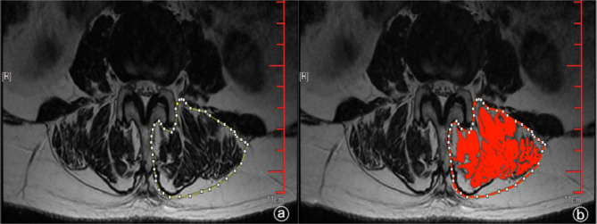
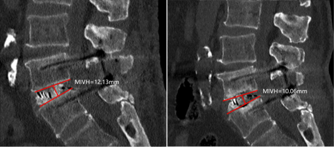
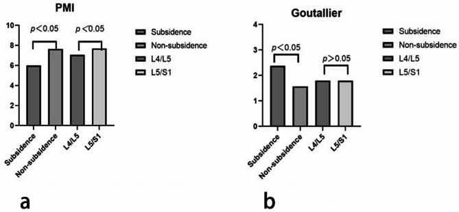
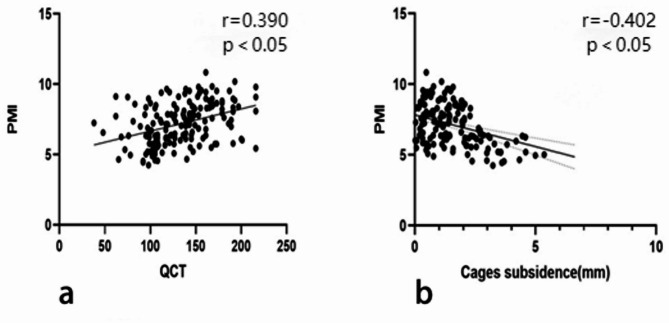
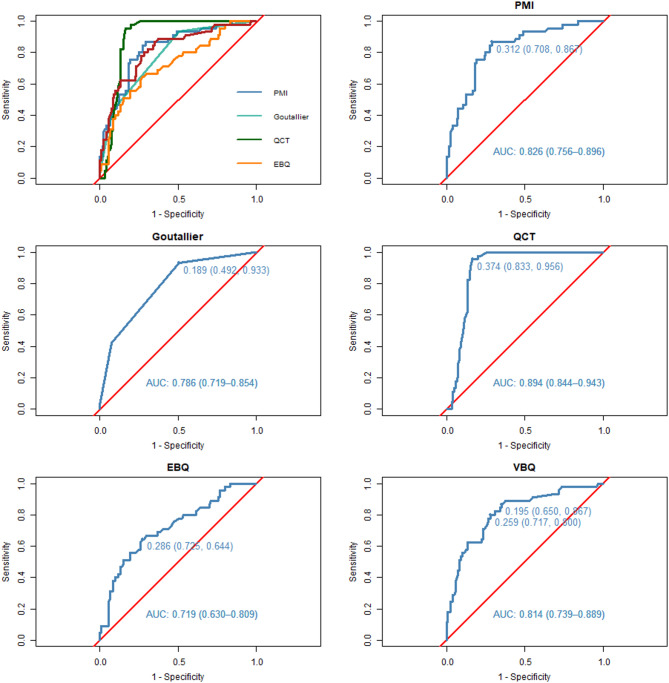
As the global population continues to age, the prevalence of lumbar degenerative disease (LDD) has increased. Meanwhile, the clinical efficacy of LDD conventional management remains limited. Posterior lumbar interbody fusion (PLIF) has become the standard surgical intervention. However, cage subsidence (CS) following PLIF poses a persistent clinical challenge. CS has been associated with several risk factors, including age, sex, low bone mineral density (BMD), endplate damage, muscle condition, and cage design. Although muscle health has recently drawn greater attention concerning surgical outcomes and BMD, dependable predictors of subsidence are still lacking. While low BMD is a recognized contributor to CS, the reliability of dual-energy X-ray absorptiometry (DEXA) is debatable. Although quantitative computed tomography (QCT) offers improved accuracy, it can be compromised by calcified structures. Similarly, fat infiltration and inflammation may affect vertebral bone quality (VBQ) and endplate bone quality (EBQ) scores. In this context, the paraspinal muscle index (PMI) and Goutallier classification (GC), both derived from magnetic resonance imaging (MRI), may serve as useful indicators of muscle quality while avoiding radiation exposure and vertebral interference. This study aimed to evaluate the predictive value of PMI and GC for CS after PLIF and compare their performance with other established imaging and bone quality markers. A retrospective review was conducted on 165 patients who underwent single-level PLIF between February 2022 and February 2024. All participants underwent preoperative MRI to assess PMI and GC and evaluate VBQ and EBQ. BMD was quantified using QCT. Patients were categorized into CS and non-CS groups based on postoperative imaging findings. Logistic regression analysis was used to identify risk factors for CS, and the predictive performance of each parameter was evaluated using receiver operating characteristic (ROC) curves, with the area under the curve (AUC) indicating diagnostic accuracy. Of the 165 patients, 45 (27.3%) developed cage subsidence. Those in the CS group were significantly older on average (70.4 ± 6.99 vs. 64.02 ± 8.24 years, p < 0.001) and had a higher proportion of female patients (p = 0.023). A lower body mass index (BMI ≤ 25 kg/m²) was less frequently observed in the CS group (p = 0.002), while no significant differences were noted for diabetes status or surgical indications. Multivariate analysis identified a lower PMI and higher GC as independent predictors of CS. ROC analysis demonstrated strong predictive performance for PMI (AUC = 0.826), GC (AUC = 0.786), QCT (AUC = 0.894), VBQ (AUC = 0.814), and EBQ (AUC = 0.719), with QCT yielding the highest diagnostic accuracy. PMI was inversely correlated with the extent of subsidence and positively associated with BMD. MRI-based assessments of muscle quality, including PMI and GC, offer reliable and non-invasive predictors of cage subsidence following PLIF. These measures may serve as practical tools in preoperative planning, enhancing risk stratification while minimizing radiation exposure.
Keywords: Cage subsidence; Goutallier classification; Lumbar degenerative disease; Magnetic resonance imaging; Paraspinal muscle index; Posterior lumbar interbody fusion.
© 2025. The Author(s).
Conflict of interest statement
Declarations. Competing interests: The authors declare no competing interests. Consent for publication: All authors had reviewed the final manuscript and gave consent for submission and publication. Ethics approval and consent to participate: This retrospective study used completely anonymized data from Yichang Central People’s Hospital. The study protocol was reviewed and approved by the Medical Ethics Committee of Yichang Central People’s Hospital (Approval No. 2023-164-01). Given the retrospective nature of the study, the requirement for informed consent was waived by the Medical Ethics Committee.
Figures
References
-
- Alentado, V. J. et al. Independent predictors of a clinically significant improvement after lumbar fusion surgery. Spine Journal: Official J. North. Am. Spine Soc.17 (2), 236–243 (2017). - PubMed
-
- Fenton-White, H. A. Trailblazing: the historical development of the posterior lumbar interbody fusion (PLIF). Spine Journal: Official J. North. Am. Spine Soc.21 (9), 1528–1541 (2021). - PubMed
-
- Amorim-Barbosa, T. et al. Risk factors for cage subsidence and clinical outcomes after transforaminal and posterior lumbar interbody fusion. Eur. J. Orthop. Surg. Traumatology: Orthopedie Traumatologie. 32 (7), 1291–1299 (2022). - PubMed
-
- Park, M. K. et al. Risk factors for cage migration and cage retropulsion following transforaminal lumbar interbody fusion. Spine Journal: Official J. North. Am. Spine Soc.19 (3), 437–447 (2019). - PubMed
MeSH terms
LinkOut - more resources
Full Text Sources
Medical
Research Materials
Miscellaneous
