

Automated Antithrombin Activity Detection with Whole Capillary Blood Based on Digital Microfluidic Platform
- PMID: 40731694
- PMCID: PMC12298285
- DOI: 10.3390/mi16070785
Automated Antithrombin Activity Detection with Whole Capillary Blood Based on Digital Microfluidic Platform
Abstract
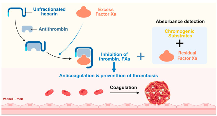
Antithrombin (AT) plays a crucial role in the human anticoagulant system and has extensive clinical applications. However, traditional detection methods often require large sample volumes, complex procedures, and lengthy processing times.
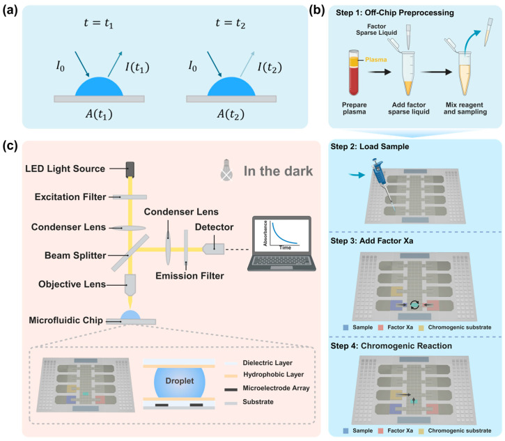
Methods: We integrated digital microfluidics technology with AT detection to develop a point-of-care testing (POCT) device that is user-friendly and fully automated for real-time AT testing.
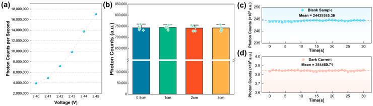
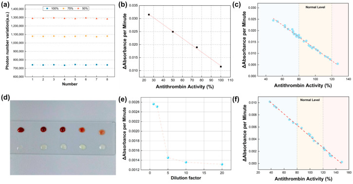
Results: This device allows for automation and enhanced adaptability to various settings, requiring only a minimal sample volume (whole capillary blood), thereby omitting steps such as plasma separation to save time and improve clinical testing efficiency. Comparisons with conventional AT activity detection methods demonstrate a high degree of consistency in the results obtained with this device.
Conclusion: The AT detection system we developed exhibits significant effectiveness and holds substantial research potential, positioning it to evolve into a clinically impactful POCT solution for AT assessment.
Keywords: AT; in vitro diagnostics; microfluidics; optical biosensors; point-of-care testing.
Conflict of interest statement
The authors declare no conflicts of interest.
Figures

Similar articles
-
Signs and symptoms to determine if a patient presenting in primary care or hospital outpatient settings has COVID-19.Cochrane Database Syst Rev. 2022 May 20;5(5):CD013665. doi: 10.1002/14651858.CD013665.pub3. Cochrane Database Syst Rev. 2022. PMID: 35593186 Free PMC article.
-
Automated monitoring compared to standard care for the early detection of sepsis in critically ill patients.Cochrane Database Syst Rev. 2018 Jun 25;6(6):CD012404. doi: 10.1002/14651858.CD012404.pub2. Cochrane Database Syst Rev. 2018. PMID: 29938790 Free PMC article.
-
Computer and mobile technology interventions for self-management in chronic obstructive pulmonary disease.Cochrane Database Syst Rev. 2017 May 23;5(5):CD011425. doi: 10.1002/14651858.CD011425.pub2. Cochrane Database Syst Rev. 2017. PMID: 28535331 Free PMC article.
-
Automated devices for identifying peripheral arterial disease in people with leg ulceration: an evidence synthesis and cost-effectiveness analysis.Health Technol Assess. 2024 Aug;28(37):1-158. doi: 10.3310/TWCG3912. Health Technol Assess. 2024. PMID: 39186036 Free PMC article.
-
What is the value of routinely testing full blood count, electrolytes and urea, and pulmonary function tests before elective surgery in patients with no apparent clinical indication and in subgroups of patients with common comorbidities: a systematic review of the clinical and cost-effective literature.Health Technol Assess. 2012 Dec;16(50):i-xvi, 1-159. doi: 10.3310/hta16500. Health Technol Assess. 2012. PMID: 23302507 Free PMC article.
References
-
- Vincent L.E., Talanker M.M., Butler D.D., Zhang X., Podbielski J.M., Wang Y.-W.W., Chen-Goodspeed A., Gonzalez S.L.H., Fox E.E., Cotton B.A., et al. Association of changes in AT activity over time with responsiveness to enoxaparin prophylaxis and risk of trauma-related venous thromboembolism. JAMA Surg. 2022;157:713–721. doi: 10.1001/jamasurg.2022.2214. - DOI - PMC - PubMed
Grants and funding
- Grants 2023YFF0721500/the National Key R&D Program of China
- YN202514/the Aerospace Center Hospital Research Fund
- Grants 62105177/the National Natural Science Foundation of China
- Grant 2023QNRC001/the Young Elite Scientists Sponsorship Program of CAST
- Grant 2024NSCQMSX3784/the Natural Science Foundation of Chongqing
LinkOut - more resources
Full Text Sources
