

Dens Invaginatus: A Comprehensive Review of Classification and Clinical Approaches
- PMID: 40731909
- PMCID: PMC12298056
- DOI: 10.3390/medicina61071281
Dens Invaginatus: A Comprehensive Review of Classification and Clinical Approaches
Abstract
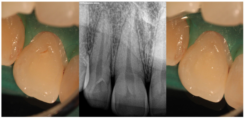
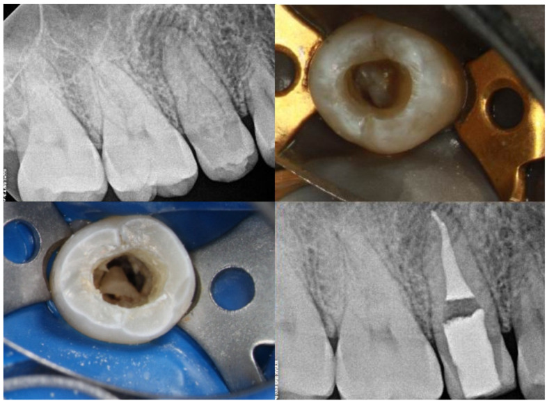
Dens invaginatus is a developmental dental anomaly characterized by the infolding of the enamel organ into the dental papilla during early odontogenesis. This process leads to a broad spectrum of anatomical variations, ranging from minor enamel-lined pits confined to the crown to deep invaginations extending through the root, occasionally communicating with periodontal or periapical tissues. The internal complexity of affected teeth presents diagnostic and therapeutic challenges, particularly in severe forms that mimic root canal systems or are associated with pulpal or periapical pathology. Maxillary lateral incisors are most frequently affected, likely due to their unique developmental timeline and morphological susceptibility. Although various classification systems have been proposed, Oehlers' classification remains the most clinically relevant due to its simplicity and correlation with treatment complexity. Recent advances in diagnostic imaging, especially cone beam computed tomography (CBCT), have revolutionized the identification and classification of these anomalies. CBCT-based adaptations of Oehlers' classification allow for the precise assessment of invagination extent and pulpal involvement, facilitating improved treatment planning. Contemporary therapeutic strategies now include calcium-silicate-based cement sealing materials, endodontic microsurgery for inaccessible anatomy, and regenerative endodontic procedures for immature teeth with necrotic pulps. Emerging developments in artificial intelligence, genetic research, and tissue engineering promise to further refine diagnostic capabilities and treatment options. Early detection remains critical to prevent complications such as pulpal necrosis or apical disease. A multidisciplinary, image-guided, and patient-centered approach is essential for optimizing clinical outcomes in cases of dens invaginatus.
Keywords: dens invaginatus; dental anomaly; endodontics; root canal therapy; treatment.
Conflict of interest statement
The authors declare no conflicts of interest.
Figures


References
-
- Versiani M.A., Martins J.N., Basrani B. The Root Canal Anatomy in Permanent Dentition. Springer; Cham, Switzerland: 2019. 3D visual glossary of terminology in root and root canal anatomy; pp. 391–425.
Publication types
MeSH terms
LinkOut - more resources
Full Text Sources
